Ultrasound strain imaging in tissue therapies

a technology of ultrasound and tissue therapy, applied in the field of radiation oncology, can solve the problems of unreliable tumor location images, inability to provide soft tissue imaging, and displace surrogate implants, so as to maximize radiation doses to tumors, reduce the exposure to surrounding healthy tissue, and maximize radiation doses

Inactive Publication Date: 2005-12-01
THE JOHN HOPKINS UNIV SCHOOL OF MEDICINE
View PDF8 Cites 16 Cited by
  • Summary
  • Abstract
  • Description
  • Claims
  • Application Information

AI Technical Summary

Benefits of technology

[0013] An advantage of the present invention is to assist in maximizing that it maximizes radiation dosages to tumors while reducing the exposure to of surrounding healthy tissue to the radiation.
[0014] Another advantage of the present invention is to that it provides more accurate imagery of a prostate the targeted anatomical structure (e.g., the prostate) during radiation therapy, which it does by taking advantage of the differences in strain properties between the targeted anatomical structure and prostate and its surrounding tissue.

Problems solved by technology

However, oOne problem with this approach is that EPIDs is that they do not provide imagery of soft tissue.
The problem with this approach is that intervening acoustic interfaces, such as the bladder, air pockets in the bladder, bone, etc., make such images unreliable for locating the tumor.
A disadvantage of such solutions is that the implanted devices are mere surrogates for the prostate, and they which do not provide information regarding that specifically defines the contours of the prostate.
In addition, these devices, and which must be invasively implanted.
Further, swelling of the prostate may occur during treatment, which could cause displace the surrogate implants to move relative to the location of the tumor.

Method used


Image

  • Ultrasound strain imaging in tissue therapies
  • Ultrasound strain imaging in tissue therapies
  • Ultrasound strain imaging in tissue therapies

Examples


Embodiment Construction

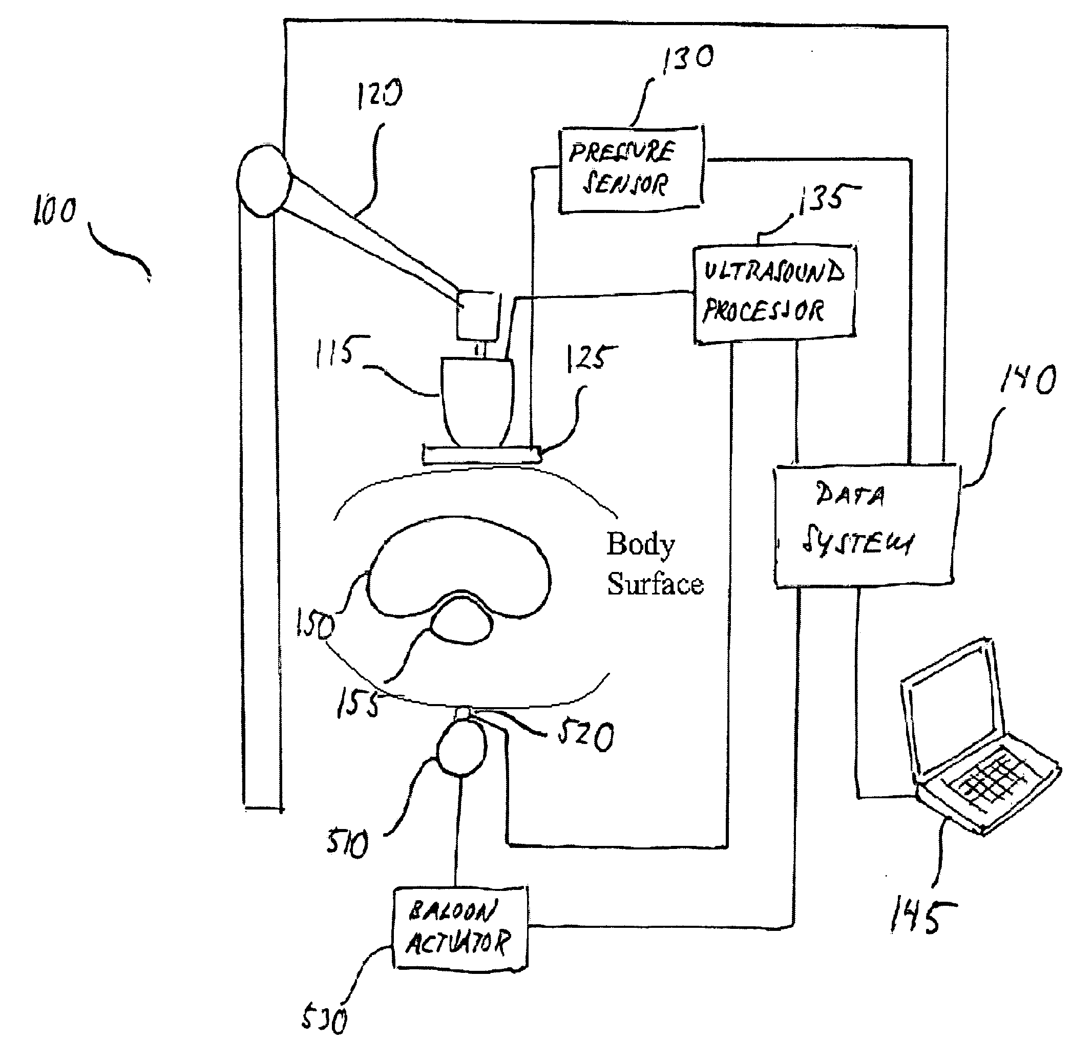
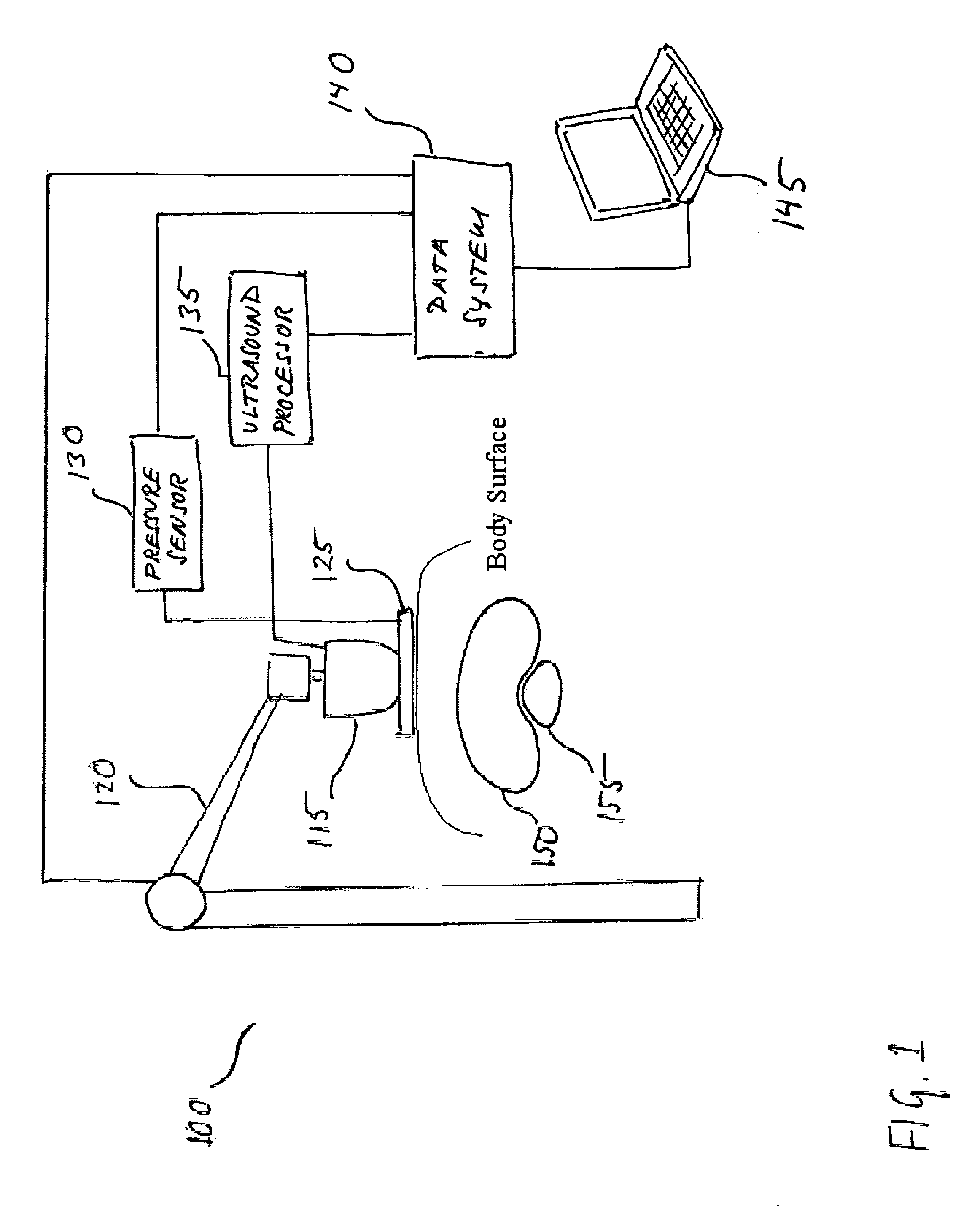
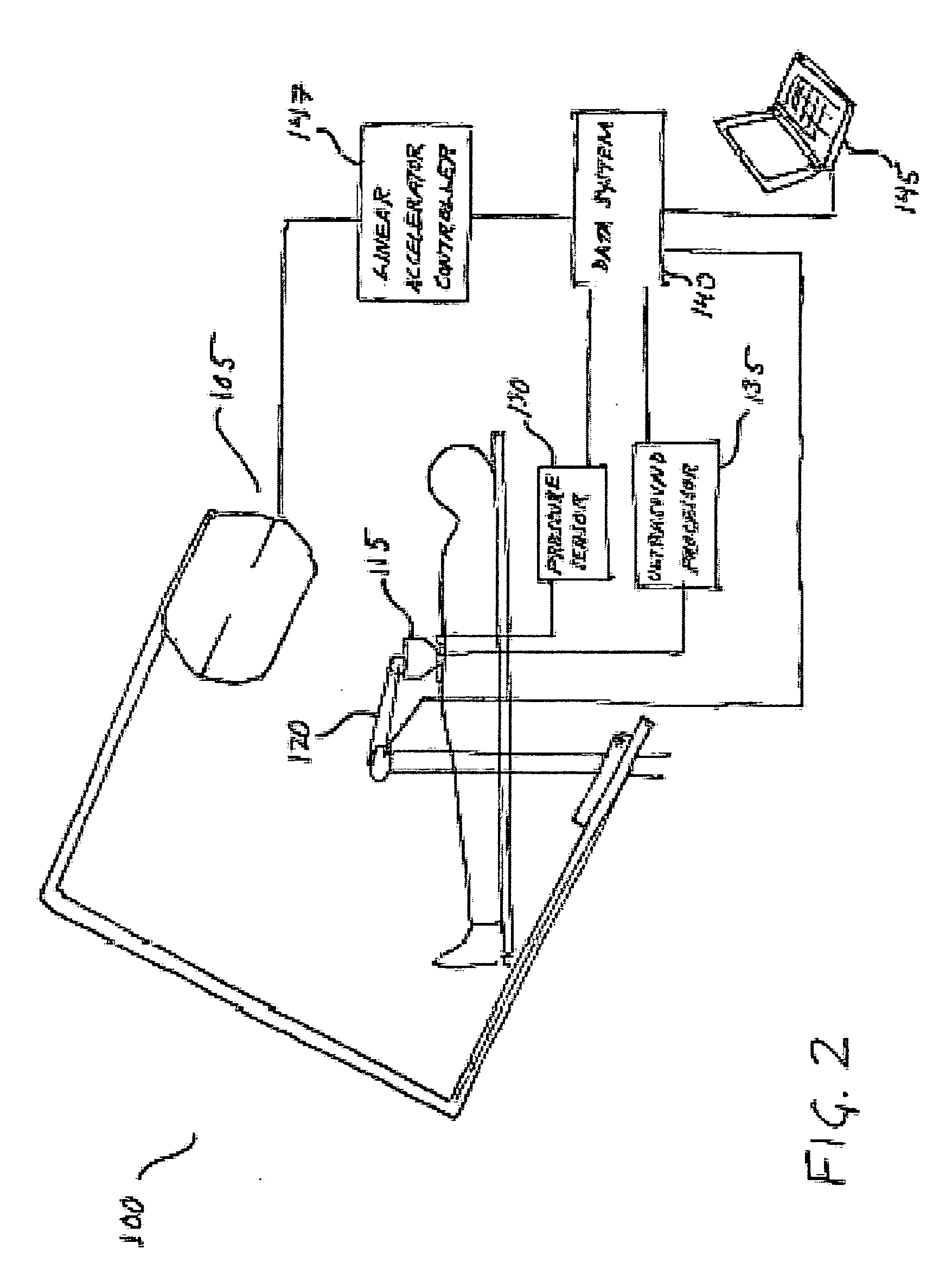
[0027] As used herein, ultrasound strain imaging refers to acquiring ultrasound imagery of an anatomical region, which includes a targeted anatomical structure and its surrounding tissue, region while precisely incrementally applying incremental amounts of substantially precise pressure against an area of patient's body in the vicinity of the anatomical region abdomen. The applied pressure exerts strain on the tissue within the abdomen anatomical region, including the targeted anatomical structure and prostate and its surrounding tissue. The surrounding tissue and the targeted anatomical structure prostate respond differently to the exerted strain such that the tissue and the targeted anatomical structure prostate will compress at different rates. By correlating the ultrasound data acquired with and without strain, the contours of the targeted anatomical structure prostate may be identified based on its different response to the strain.

[0028] For purposes of illustration and not li...


PUM


Abstract

Disclosed is a system and method for providing ultrasound strain imaging of a soft tissue, such as a prostate, so that the soft tissue may be more precisely targeted during radiation treatment. The system includes a mechanical arm with a pressure interface for applying incremental pressure to the patient's abdomen, and a data system for correlating the ultrasound signals acquired before and after each application of incremental pressure. By correlating the pre and post-pressure ultrasound signals, acoustic interfaces corresponding to the prostate, which define the contours of the prostate, may be displayed. Further, data corresponding to the contours of the prostate may be provided to a linear accelerator to enable the linear accelerator to more precisely target the prostate.

Description

[0001] This application claims the benefit of U.S. Provisional Patent Application No. 60 / 569,003, filed on May 7, 2004; U.S. Provisional Patent Application No. 60 / 577,789, filed on Jun. 8, 2004; and U.S. Non-provisional Application Ser. No. 10 / 895,397 titled ROBOTIC 5D ULTRASOUND SYSTEM, which claims priority to U.S. Provisional Patent Application No. 60 / 488,941, filed Jul. 21, 2003, all of which are hereby incorporated by reference for all purposes as if fully set forth herein.[0002] The research and development effort associated with the subject matter of this patent application was supported by the National Science Foundation under grant no. EEC9731478. BACKGROUND OF THE INVENTION [0003] 1. Field of the Invention [0004] The present invention relates primarily to the field of radiation oncology. More particularly, the present invention involves a system and method to provide more precise targeting of radiation by improving ultrasound imaging of the target tissue. [0005] 2. Discuss...

Claims


Application Information

Patent Timeline
Patent Type & Authority Applications(United States)
IPC IPC(8): A61B8/00A61B8/08A61B17/00A61B19/00
CPCA61B5/0048A61B5/6843A61B8/08A61B8/0833A61B2018/00547A61B8/485A61B19/26A61B2017/00274A61B8/4218A61B8/4281A61B90/50
Inventor BOCTOR, EMAD M.FICHTINGER, GABOR
Owner THE JOHN HOPKINS UNIV SCHOOL OF MEDICINE
Why Eureka
Try Eureka