Methods and compositions for bioengineering a tooth
a technology of tooth tissue and composition, applied in the field of methods and compositions for bioengineering tooth tissue, can solve the problems of tooth loss in aged populations, significant incidence of children born with missing primary and/or adult teeth, and high incidence of tooth loss in children
- Summary
- Abstract
- Description
- Claims
- Application Information
AI Technical Summary
Benefits of technology
Problems solved by technology
Method used
Image
Examples
example 1
Identification and Cloning of Dental Precursor Cells
[0032] Expression of STRO-1 in Cultured Epithelial and Mesenchymal Dental Cells
[0033] Tissue samples were taken from pig and rat post-natal tooth buds. The tissue samples were minced to create tooth bud cell suspensions. Optionally, the minced tissue fragments are further subject to enzymatic dissociation, e.g., using collagenase, to produce single cell suspensions. The cell suspensions were cultured using standard cell culture techniques.
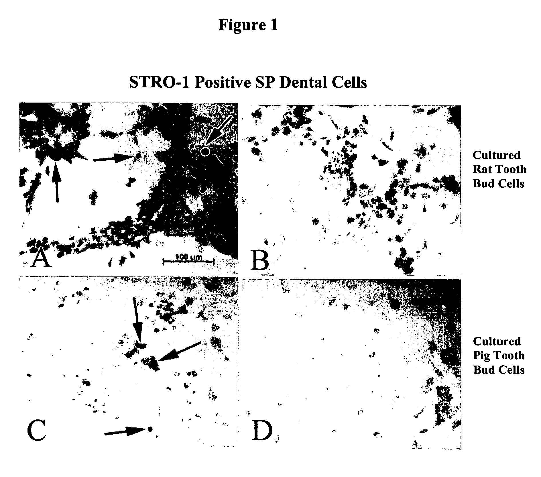
[0034] Immunohistochemical analysis of the cultured pig and rat tooth bud cells was performed using a monoclonal antibody to STRO-1 (The Developmental Studies Hybridoma Bank, Iowa City, Iowa), a surface marker for bone marrow stem cells. The presence of STRO-1 positive (STRO-1+) cells was used a marker for identifying dental precursor cells in the cultured tooth bud cells. The results of this immunohistochemical analysis are shown in FIGS. 1A-1D, where FIG. 1A depicts the expression of STRO-1 i...
example 2
Biocompatible Implants
[0042] Cultured pig tooth bud cells were seeded onto a scaffold at a cell density of 7.0×10⁵ cells / mm3. The cell / scaffold construct was then implanted at a highly vascularized site, the omentum, of a mammalian host. Omentum surgeries were performed as previously described. (Young et al., J. Dent. Res., 81:695-700 (2002)). Implantation resulted in the formation of mixed bioengineered dentin and enamel tissues that approximated the size of the scaffold (FIG. 4). After a period of time, e.g., 18-20 weeks post-implantation, mature tooth tissue and de novo tooth development was observed. De novo tooth development after implantation of the cell / scaffold construct was used as another marker of dental precursor cells. Detection of de novo tooth development throughout the life of the omental implant suggests the in vivo presence and maintenance of these dental precursor cells.
example 3
Detection of Dentin / Enamel Junction (DEJ) Formation
[0043] Enamel organ and pulp organ tissues were isolated and used to generate single cell suspensions of dental epithelial or mesenchymal cells, respectively, as described above in Example 1. A first scaffold was seeded either with 1.5×10⁷ enamel epithelial cells, and a second scaffold was seeded with 1.0×10⁷ pulp mesenchymal cells. These scaffolds were sutured together, implanted in the omentum of nude rat hosts, and grown for 8 weeks. Histological analyses of harvested implant tissue revealed adjacent layers of dentin and enamel that aligned with the interface of the two scaffolds (FIG. 5, Panel A, scaffold interface is indicated by red dotted line). No scaffold-guided DEJs were observed in scaffolds seeded with mixed epithelial and mesenchymal dental cell populations. In addition, scaffold implants seeded with dental mesenchymal cells alone formed osteodentin, and implants seeded with dental epithelial cells alone did not form e...
PUM
| Property | Measurement | Unit |
|---|---|---|
| Composition | aaaaa | aaaaa |
| Level | aaaaa | aaaaa |
| Biocompatibility | aaaaa | aaaaa |
Abstract
Description
Claims
Application Information
 - R&D
- Intellectual Property
- Life Sciences
- Materials
- Tech Scout
- Unparalleled Data Quality
- Higher Quality Content
- 60% Fewer Hallucinations
Browse by: Latest US Patents, China's latest patents, Technical Efficacy Thesaurus, Application Domain, Technology Topic, Popular Technical Reports.
© 2025 PatSnap. All rights reserved.Legal|Privacy policy|Modern Slavery Act Transparency Statement|Sitemap|About US| Contact US: help@patsnap.com


