Live cell biosensors

a biosensor and live cell technology, applied in the field of environmental sensitive dyes, can solve the problems of low sensitivity, preventing the study of many important targets, and limiting the use of refret-based techniques,

Inactive Publication Date: 2006-02-09
THE SCRIPPS RES INST
View PDF11 Cites 18 Cited by
  • Summary
  • Abstract
  • Description
  • Claims
  • Application Information

AI Technical Summary

Benefits of technology

[0016] wherein each X separately represents a variable amino acid, n is an integer between about 3 and ...

Problems solved by technology

These approaches can be useful, but FRET-based techniques suffer from limitations that prevent the study of many important targets.
Proteins undergoing conformational changes often cannot be “sampled” by a biosensor because the protein is bound to a competing ligand or is incorporated in a multi-protein complex, where it is blocked from biosensor acc...

Method used


Image

  • Live cell biosensors
  • Live cell biosensors
  • Live cell biosensors

Examples


example 1

Synthesis of Dyes

[0145] This Example describes the synthesis of useful, water-soluble derivatives of the 1a dye. New analogues were made, for example, by substitution of a quaternary amino group in the 1a dye with a much more hydrophilic sulfonato group (—SO3Na). In some analogues, cysteine-selective iodoacetamido groups were also added (e.g., at R2) for covalent attachment to binding domains.

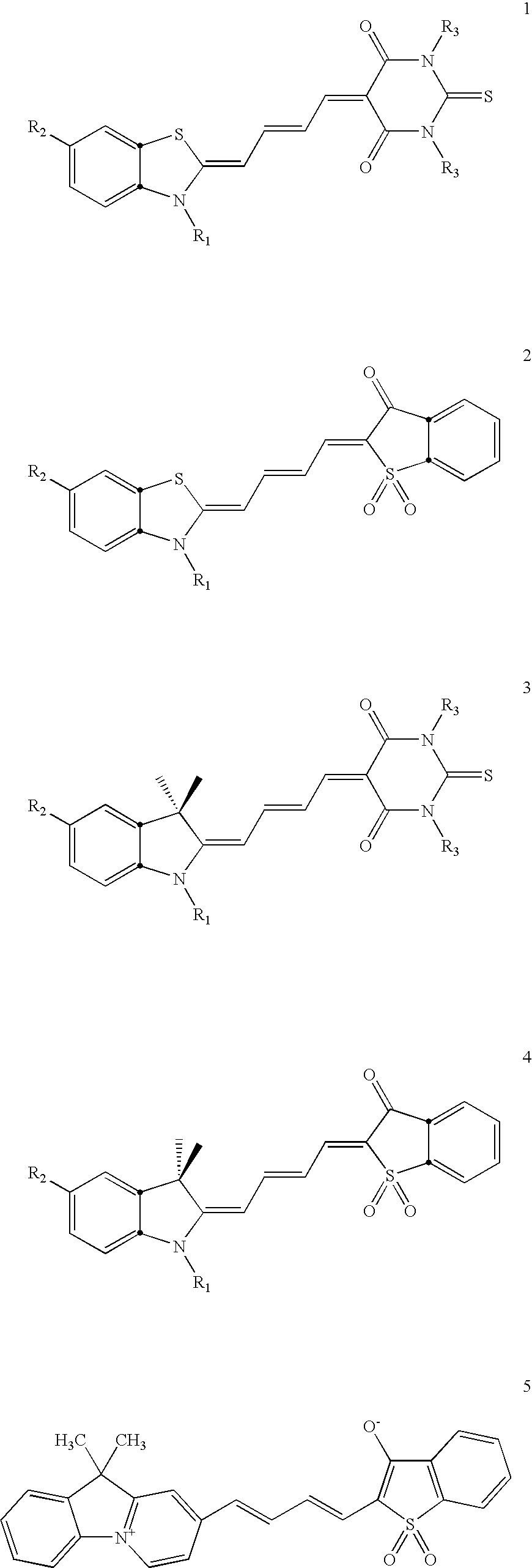
[0146] Dyes having formulae 1-4 were synthesized. Formula 1 is as follows.

[0147] Structure 1 is also referred to herein as S-TBA. A number of different dye compounds were made that had formula 1, including those with structures 1a-1h. The substituents present on each of dye compounds 1a-1h were as follows:

R1═—(CH2)3—N+(CH3)2—(CH2)2—NCS; R2═H; R3=butyl.   1a

R1=ethyl; R2═H; R3=methyl.   1b

R1═—(CH2)3—SO3−; R2═H; R3=methyl.   1c

R1═—(CH2)3—SO3−; R2=FmocNH—; R3=methyl.   1d

R1═—(CH2)3—SO3−; R2═NH2; R3=methyl.   1e

(S-TBA-IAA): R1═—(CH2)3—SO3−; R2═ICH2CONH2; R3=methyl.   1f

R1═—(CH2)3—SO3−; R2...

example 2

Methods for Making and Using Biosensors

[0251] This Example illustrates how to make and use the biosensors of the invention. The specific biosensor described here was based on the HIV-1 neutralizing antibody Fab fragment X5, which binds to HIV envelope protein gp120 after forming a complex with the host cell receptor CD4. See Moulard, M. et al., Broadly Cross-reactive HIV-1-Neutralizing Human Monoclonal Fab Selected for Binding to gp120-CD4-CCR5 Complexes. Proc Natl Acad Sci USA 99, 6913-6918 (2002).

Materials

[0252] The SB medium and phagemid pComb3X employed were obtained as described in Barbas et al., Phage Display: A Laboratory Manual. (Cold Spring Harbor Laboratory Press, Cold Spring Harbor; 2001). Soluble CD4 and IgG 2G12 were obtained from the National Institute of Health AIDS Research and Reference Program (ARRRP); soluble CD4 was also obtained from Progenics Pharmaceuticals, Inc. Gp120BaL was obtained from Quality Biological Inc. (HIV BaL rgp120).

Recombinant DNA Procedur...

example 3

Detecting Phosphorylation Induced Changes in Erk

[0284] This Example illustrates that the dyes of the invention can be attached directly to a protein of interest (e.g. Erk), where the dyes respond to conformational changes and phosphorylation. In particular, the I-TBA-3CNPh dye was attached to cysteine 214 of the MAP kinase Erk2, where it responded to Erk phosphorylation. A structure for the I-TBA-3CNPh dye is provided below.

[0285]FIG. 15A shows the I-TBA-3CNPh dye in a space filling model as it is thought to fit into the Erk2 structure. This model was based on tryptic digestion combined with mass spectroscopic sequencing of the labeled protein, and on docking studies. The lighter (yellow) residues represent two lysines undergoing hydrogen bonding with the dye, and a tryptophan interacting through π-π stacking. Phosphorylation of the purple residues altered the conformation of the protein, affecting the tryptophan interaction and perturbing some of the hydrogen bonds. Such phospho...


PUM

PropertyMeasurementUnit
Fractionaaaaaaaaaa
Fractionaaaaaaaaaa
Wavelengthaaaaaaaaaa

Abstract

The invention provides dyes, biosensors, and methods for using the dyes and biosensors to detect selected target molecules. The biosensors have a binding domain and a dye, or a dye which is attached directly to the target of interest. Binding domains contemplated by the invention include biomolecules or fragments of biomolecules that interact with target molecules of interest and can be specific to a given conformational state or covalent modification of the molecule (e.g. phosphorylation). In one embodiment, the binding domain of a biosensor is a single chain variable fragment (scFv) with a dye of the invention linked to a CDR3 region. The invention also provides environmentally sensitive dyes useful for detecting changes in the binding, conformational change, or posttranslational modification of the selected target.

Description

[0001] This application claims benefit of the filing date of U.S. Provisional Application Ser. No. 60 / 552,663, filed Mar. 12, 2004, the contents of which are incorporated herein in their entirety.GOVERNMENT FUNDING [0002] The invention described herein was made with United States Government support under Grant Numbers R01-AG-15430 and R01-GM-57464 awarded by the National Institutes of Health. The United States Government has certain rights in this invention.FIELD OF THE INVENTION [0003] The invention relates to environmentally-sensitive dyes, use of the dyes to report protein activities, biosensor molecules that can bind to selected targets, and methods for detecting target biomolecules and protein activities, for example, within living cells. BACKGROUND OF THE INVENTION [0004] Cell behavior is regulated through transient activation of protein activities at specific subcellular locations. Our ability to study translocation of proteins has been greatly enhanced by advances in the mic...

Claims


Application Information

Patent Timeline
IPC IPC(8): C12Q1/68G01N33/53C12M1/34C07K1/107C07K1/13C12N15/82G01N33/532G01N33/542G01N33/58
CPCC07D403/06C07D409/06C07D417/06C07D471/04C07K2319/60G01N2500/10G01N33/573G01N33/582G01N33/6857G01N2333/16G01N2333/916G01N33/542Y10S436/80
Inventor HAHN, KLAUS
Owner THE SCRIPPS RES INST
Why Eureka
Try Eureka