

Method for vitrification of mammalian cells
a technology for vitrification and mammalian cells, applied in the field of vitrification of mammalian biological specimens, can solve the problems of poor survival and development rate, and low development rate of cryopreservation techniques in most species, and achieves low pregnancy rate, poor recovery of vitrified specimens, and convenient use.
- Summary
- Abstract
- Description
- Claims
- Application Information
AI Technical Summary
Benefits of technology
Problems solved by technology
Method used
Image

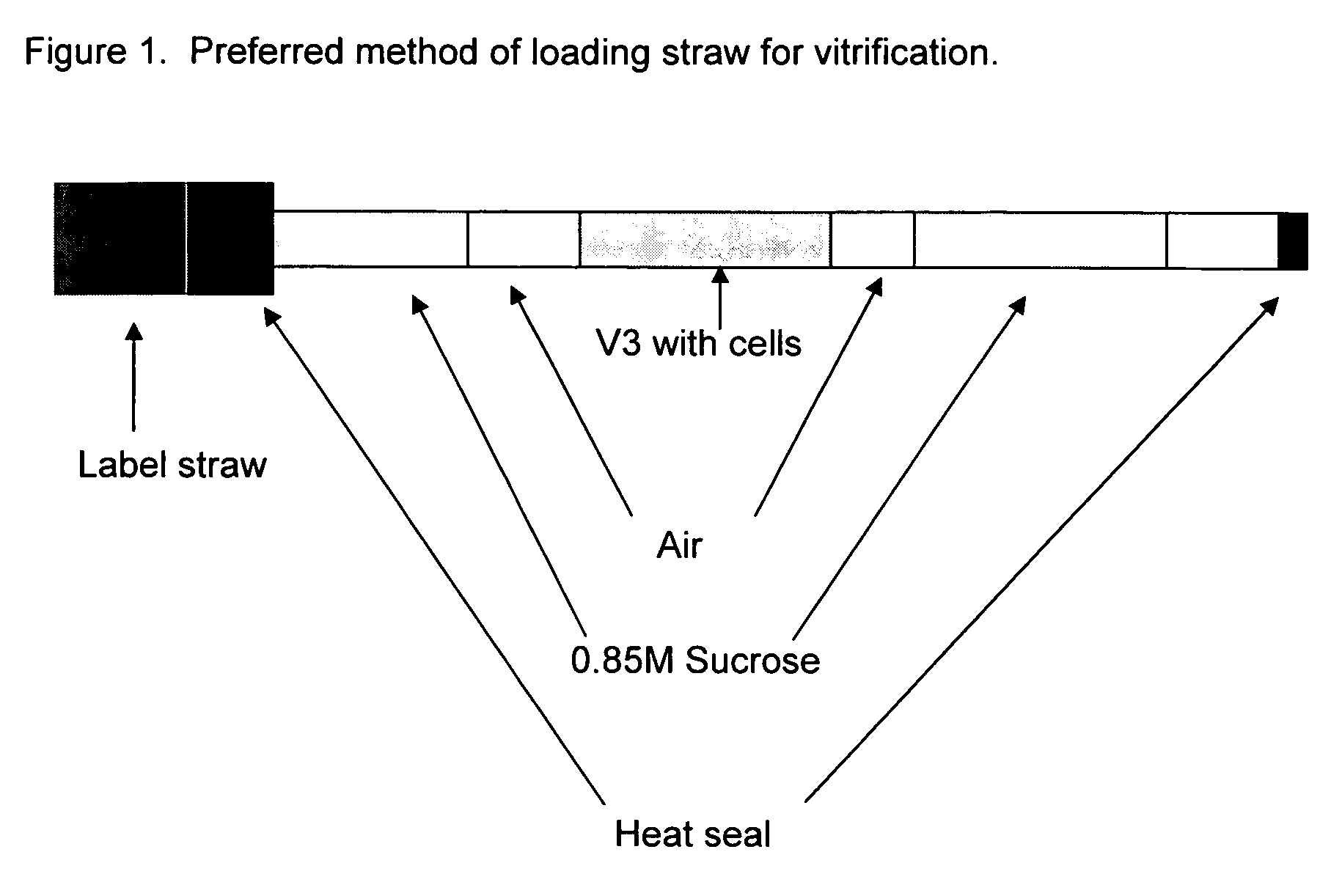
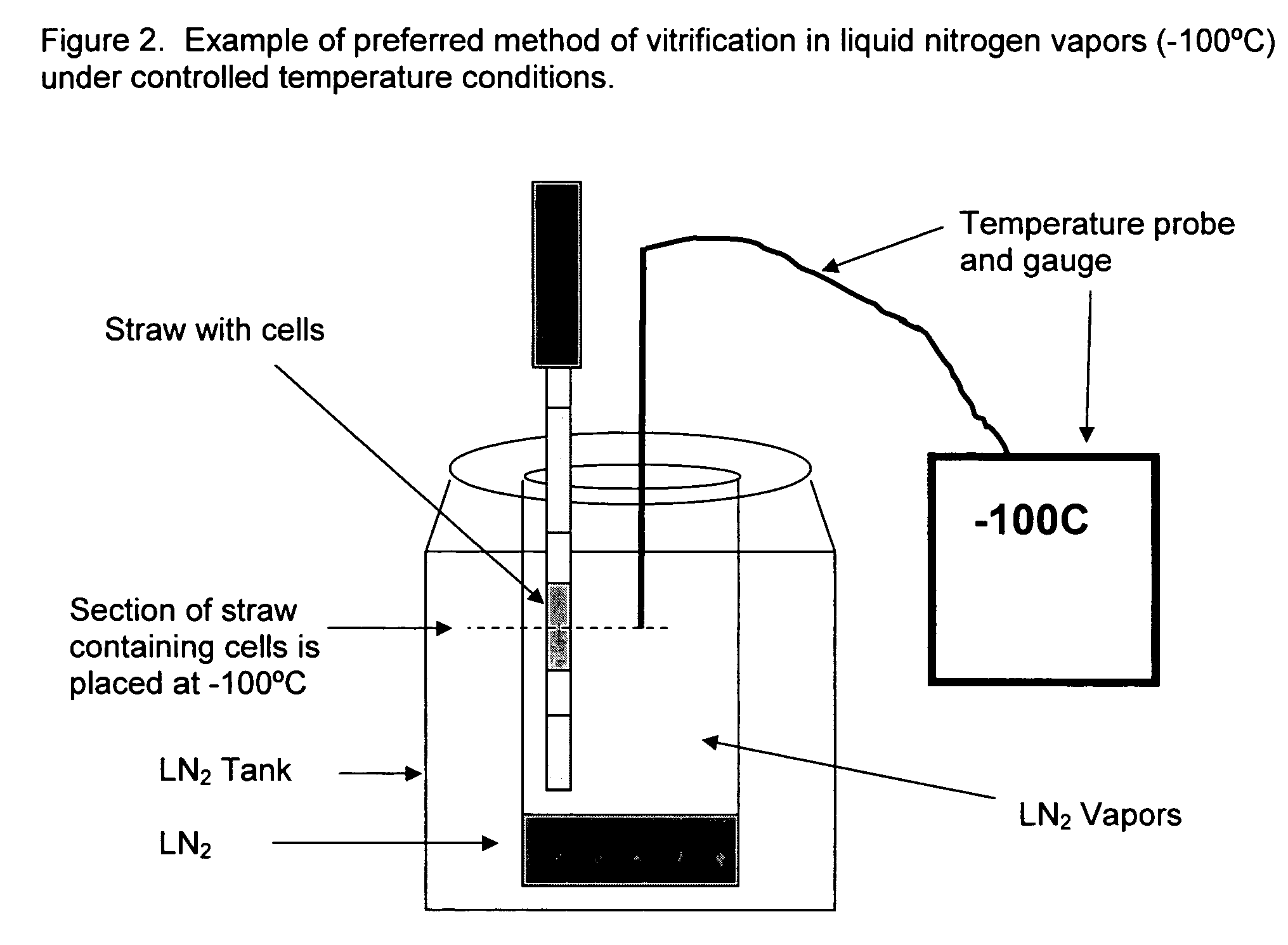
Examples
example 1
Vitrification of Bovine and Human Blastocysts
[0056] All human embryos were discarded material donated to research. For all embryos used, written consent was obtained from patients in accordance with each internal review board protocol. A human embryo on Day 5, 6, or 7 that had a visible blastocoel was designated as a blastocyst and vitrified. Most of the embryos had questionable inner cell mass quality, or poorly defined trophoblast and inner cell mass cells.
[0057] Bovine oocytes were purchased from BoMed (Madison, Wis.) and shipped overnight in a portable heated incubator. Oocytes were cultured for several hours in order for full maturation to occur (24 h from start of culture) before insemination with bull sperm. Oocytes were inseminated in IVF-TALP. After incubation with sperm overnight, the oocytes were washed and cultured in cSOF, supplemented with essential and non-essential amino acids (Gibco BRL). After 5 days of culture, good quality embryos (16-cell to Morulae) were tran...
PUM
 Abstract
Description
Claims
Application Information