
Method for two-dimensional conformation-dependent separation of non-circular nucleic acids

a two-dimensional conformation-dependent separation and non-circular nucleic acid technology, applied in the field of nucleic acid analysis, can solve the problems of limited use of previously described methods for conformation-dependent separation of linear dna fragments, such as ha and csge, and significant conformational changes of the entire molecul

Inactive Publication Date: 2006-03-16
LIFEIND EHF
View PDF1 Cites 6 Cited by
  • Summary
  • Abstract
  • Description
  • Claims
  • Application Information

AI Technical Summary

Benefits of technology

[0014] Methods are provided for, conformation-dependent separation of complex mixture of linear nucleic acid fragments independent of their length. Methods of the present invention can be applied to linear nucleic acid fragments obtained from different sources and they do not require any special prior manipulation of the nucleic acid fragments. The present invention provides general methods that can be used in different contexts such as but not limited to; physical separation of mismatched heterohybrids from perfectly matched hetero- and homohybrids allowing isolation and enrichment of either class; simultaneous scanning of multiple fragments for mutations; isolation of damaged DNA molecules from undamaged molecules; and estimation of efficiency of nucleic acid reannealing.

Problems solved by technology

Unusual secondary structures in linear DNA fragments often lead to significant conformational changes of the entire molecule.
Use of previously described methods for conformation-dependent separation of linear DNA fragments, such as HA and CSGE, are limited to situations were the analyzed DNA molecules are of known length.
If the sample contains a complex mixture of DNA fragments of different lengths it would be impossible to identify which of them show difference in migration because of their conformation since individual bands would overlap or not resolve sufficiently.
This is a major drawback since it limits application of this technology.
Similar limitations would also apply to other comparable techniques based on capillary electrophoresis or chromatography (e.g. dHPLC).
In our experience, it is not possible to separate mismatched duplexes from perfectly matched duplexes using temperature as a variable between the two dimensions.
The combined agarose-PAGE system is limited by difficulties in transferring linear DNA fragments between the two different matrixes in an efficient and reproducible manner.
It should be emphasized that this method cannot be used to separate linear DNA fragments according to conformation because of the fact that linear DNA fragment of same length with different conformation are separated almost entirely according their size in agarose gel electrophoresis.
Further, perhaps due to these limitations, the reference does not disclose or suggest 2-D gel electrophoresis methods for separating other types of conformational different DNA molecules, such as curved linear DNA fragments containing unusual secondary structure, e.g. mismatched duplexes.
The difference in conformation of such linear DNA fragment with or without unusual local secondary structures is not great enough to ensure different migration in agarose elctrophoresis and will therefore not be detected in this system.
Such transfer often gives rise to trailing effects, which lower the detection capability of the system.
This separation of base pairs and concurrent untwisting results in increased length and stiffness of the DNA molecule thus affecting its whole conformation [10].

Method used


Image

  • Method for two-dimensional conformation-dependent separation of non-circular nucleic acids
  • Method for two-dimensional conformation-dependent separation of non-circular nucleic acids
  • Method for two-dimensional conformation-dependent separation of non-circular nucleic acids

Examples


example 1

Separation and Detection of Cy5 Labeled Linear DNA Fragments Containing Cytosine Bulges from Perfectly Matched Linear DNA Fragments Using Intercalator Approach.

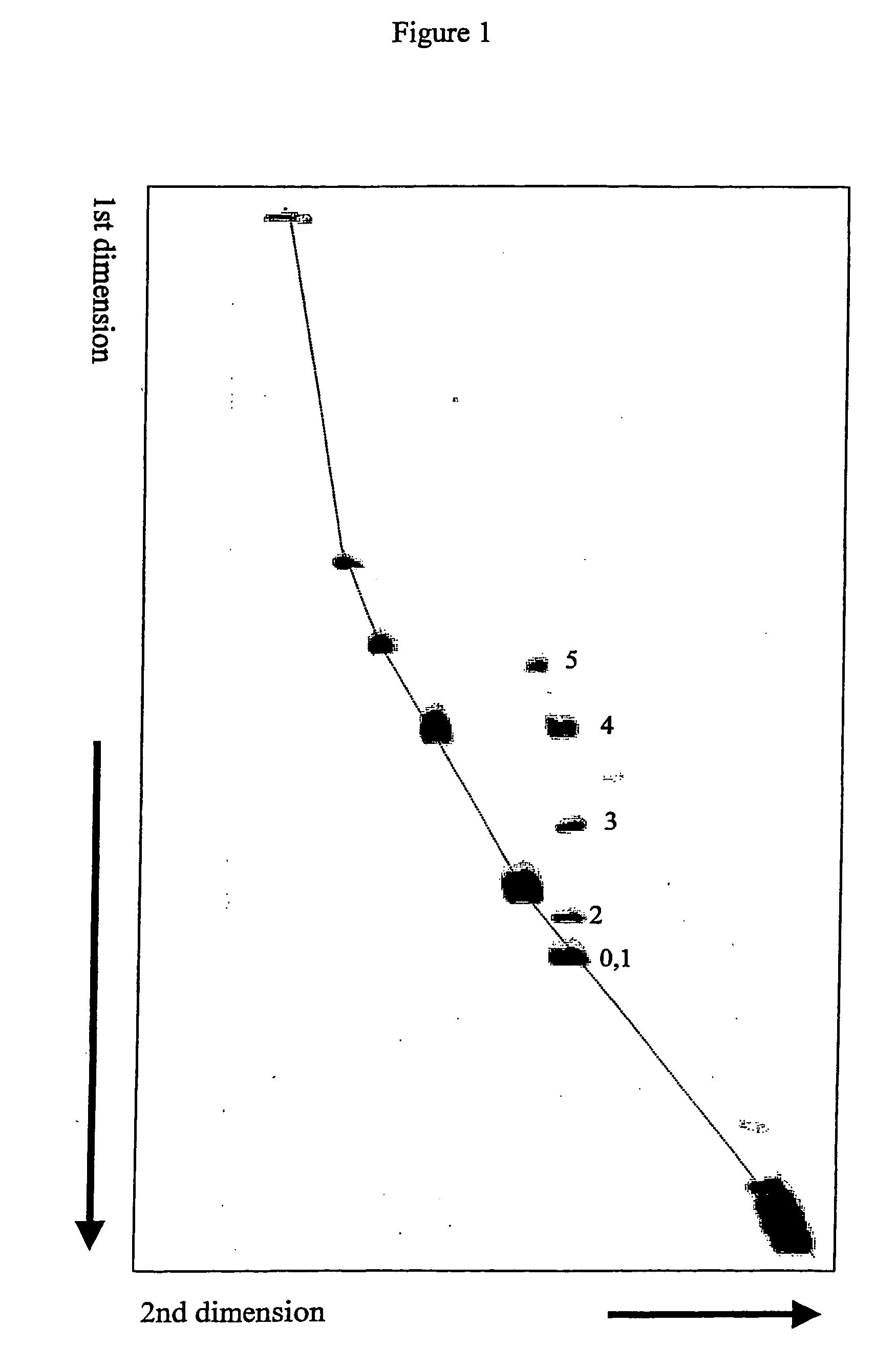
[0068] Five diffrent 298 bp linear DNA fragments were prepared. Each fragment contained one defined cytosine bulge at its center with the bulge size ranging from 1 to 5 cytosines. Otherwise the DNA fragments were identical to each other. A perfectly matched 298 bp linear DNA fragment was also prepared. Execpt for not having a cytosine bulge this fragment had identical base sequence compared to the bulged DNA fragments. These 298 bp linear DNA fragments were prepared using three shorter DNA fragments. Two fragments were PCR amplified from the plasmid pBR322. One fragment (127 bp) was 5′ end-labeled with Cy5 fluorescent molecule and contained 3′ asymmetric overhang after digestion with Ava I. The other (132 bp) contained 5′ asymmetric overhang after digestion with Ban II. One fragment (31 bp) was synthesized as two oligonucle...

example 2

Separation and Detection of Linear DNA Fragments Containing Cytosine Bulges at their Center or Near One End of the Fragment from Perfectly Matched DNA Fragments Using Intercalator Approach.

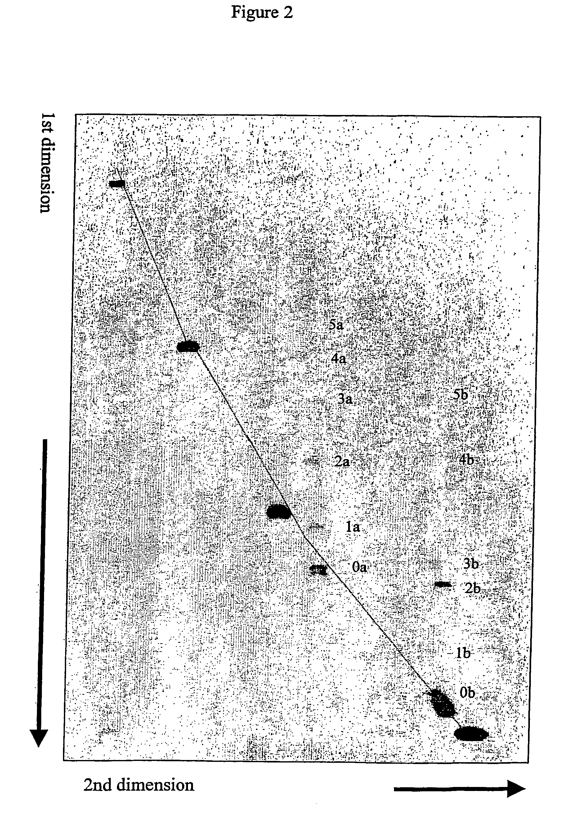
[0073] Unlabeled linear DNA fragments containing defined cytosine bulges were prepared as described in Example 1. Instead of doing equimolar ligation, ligation with excess of one end fragment was done resulting in formation of two major ligation products. One was the 298 bp product with the bulge at the center as described in Example 1. The other was a 158 bp product formed by ligation between the 31 bp synthetic molecule containing the bulge molecule and the 127 bp PCR fragment. This fragment has the bulge 15 bp from its end.

[0074] Five perfectly matched DNA fragments (155 bp, 357 bp, 543 bp, 857 bp and 1395 bp) containing 5′ Cy5 label were also prepared using PCR amplification from the plasmid pBR322.

[0075] A mixture of the fragments described above was separated by 2-D CDE gel electrophores...

example 3

Separation and Detection of Cy5-Labeled Linear DNA Fragments Containing Cytosine Bulges from Perfectly Matched Fluorescein Labeled DNA Fragments in 7×8 cm Gel Format Using Intercalator Approach.

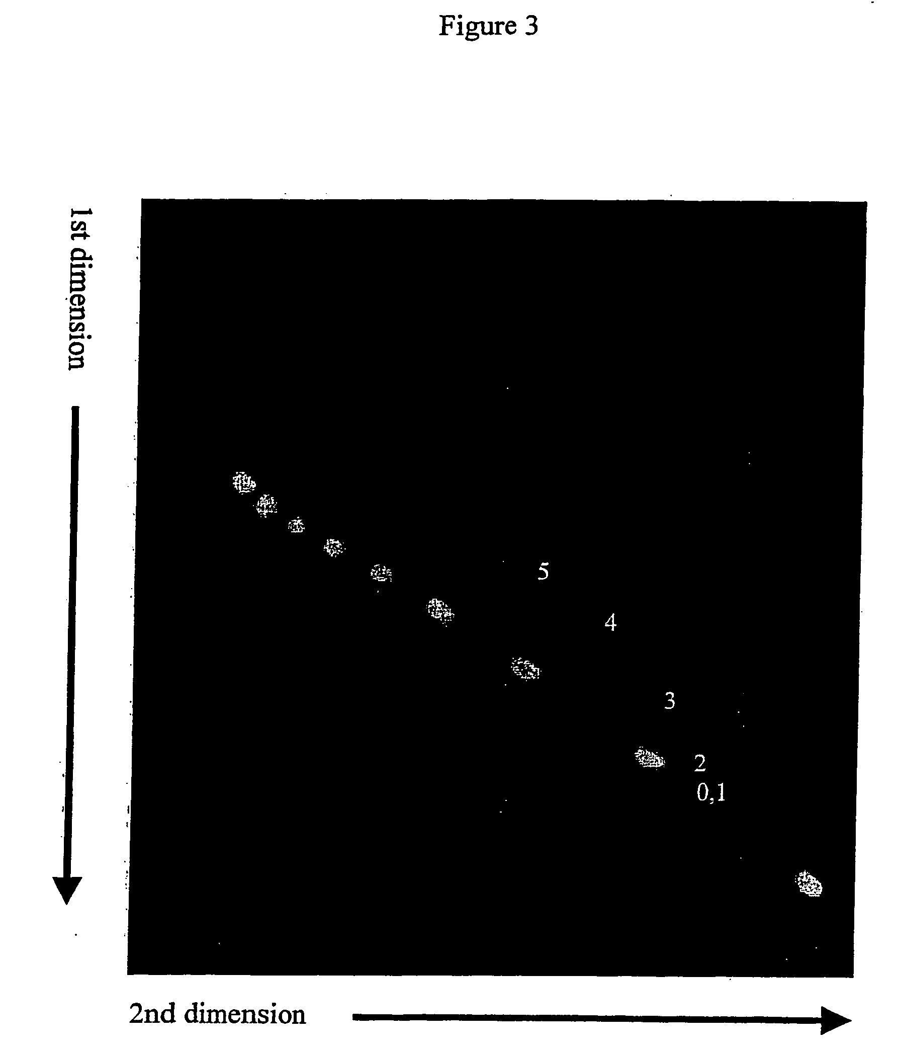
[0078] Linear Cy5-labeled DNA fragments containing cytosine bulges were prepared as described in Example 1. These DNA fragments were combined with 100 bp fluorescein ladder (BioRAD) containing 100, 200, 300, 400, 500, 600, 700, 800, 900 and 1000 bp DNA fragments. This DNA mixture was then separated by 2-D gel electrophoresis using 6% polyacrylamide gel. The gel was polymerized in 1×TBE buffer (89 mM Tris base, 89 mM borate, and 2 mM EDTA) for 1 hour. The first dimension gel electrophoresis was done in BioRad Mini Protean II vertical electrophoresis system using 7×8 cm glass sandwich with 1 mm spacers. The gel was run at room temperature for 90 minutes at 20 mA with 1×TBE in both upper and lower buffer chambers. The gel was removed from the glass sandwich and soaked in 100 ml of 1×TBE buffer...


PUM

PropertyMeasurementUnit
temperatureaaaaaaaaaa
anglesaaaaaaaaaa

Abstract

Methods are provided for separating linear nucleic acid fragments based on their conformation. The methods are based on novel two-dimensional gel electrophoresis techniques. In one aspect the method comprises electrophoresing a sample of nucleic acids in a first dimension said sample through a gel matrix under a first set of pre-determined electrophoresis conditions; electrophoresing said gel matrix in a second dimension under a second set of electrophoresis conditions, such that linear nucleic acid fragments of equal length but having different conformation are separated; said first and second electrophoresis conditions are different, such that in one of said dimensions electrophoresis allows separation of linear nucleic acid fragments based on conformation and length, and in the other of said dimensions electrophoresis allows separation of the sample fragments based substantially on length. Said difference is preferably established with a chemical agent capable of reducing conformational differences between linear nucleic acids fragments.

Description

FIELD OF THE INVENTION [0001] The present invention is in the field of nucleic acid analysis, specifically for screening nucleic acid preparations to identify, isolate, and characterize linear nucleic acid fragments with altered conformation. Examples include, but are not limited to, conformational alterations due to DNA polymorphism and modifications in structure of bases or backbone. Methods of the invention are especially useful for separation and isolation of non-circular mismatched DNA fragments from a complex mixture of hetero- and homohybrids based on their differences on conformation. BACKGROUND OF THE INVENTION [0002] Unusual secondary structures in linear DNA fragments often lead to significant conformational changes of the entire molecule. Such conformational changes of linear nucleic acids fragments frequently result in curvature, characterized by decreased end-to-end distance. Curvature has for example been detected or proposed in linear DNA fragments containing mispair...

Claims


Application Information

Patent Timeline
Patent Type & Authority Applications(United States)
IPC IPC(8): C12Q1/68C12N15/09C12N15/10
CPCC12N15/101
Inventor JONSSON, JONTHORMAR, HANSGUNNARSSON, GUDMUNDURGUDMUNDSSON, BJARKI
Owner LIFEIND EHF
Learn More