Method and system of obtaining improved data in perfusion measurements

a technology of perfusion measurement and data acquisition, applied in the field of methods and systems of obtaining improved data in blood perfusion measurement, can solve the problems of inability to evaluate the perfusion process, inability to realize real-time processes, and overestimation of the mit and the underestimation of the bf, so as to reduce the effect of partial voluming (pv) effect and remove contrast recirculation

Active Publication Date: 2006-04-20
APOLLO MEDICAL IMAGING TECH
View PDF28 Cites 21 Cited by
  • Summary
  • Abstract
  • Description
  • Claims
  • Application Information

AI Technical Summary

Benefits of technology

[0056] The program means may further direct the processor to determine a venous input function (VIFa(t)) from a draining vein to estimate an AIFa(t) where a selected artery has partial voluming, the vein being larger than the artery.
[0057] The program means may further direct the processor to determine the profile of a venous input function (VIFa(t)) from a large draining vein. The AIFa(t) may then be scaled up to have the same first-pass bolus peak area as the VIFa(t) to minimize partial voluming (PV) effect from the AIFa(t). The first-pass AIFa(t) and VIFa(t) profiles can be obtained by fitting the profiles to gamma-variate function (GVF) profiles respectively to remove contrast recirculation effects.

Problems solved by technology

However, in the case of major vessel disease, such as acute stroke or carotid artery stenosis, the measured AIF is often associated with a delay and dispersion before it reaches the tissue of interest, and causing overestimation of the MIT and underestimation of the BF.
However results of the perfusion process may not be evaluated until some time after the initial injection of the contrast agent and is thus not a real time process.
Furthermore there is no disclosure of obtaining quantitative data relating to blood flow and blood volume which can assist a physician to make a relatively quick and accurate diagnosis and decide on what steps can be taken to treat the patient.
More particularly this document does not account for any delay or dispersion of the contrast agent in an initial bolus injection.
A disadvantage of measuring the signal change in arteries using MRI is that it does not provide a true indication of the contrast volume as MRI depends upon electromagnetic fields that are altered due to the contrast agent.

Method used


Image

  • Method and system of obtaining improved data in perfusion measurements
  • Method and system of obtaining improved data in perfusion measurements
  • Method and system of obtaining improved data in perfusion measurements

Examples


Embodiment Construction

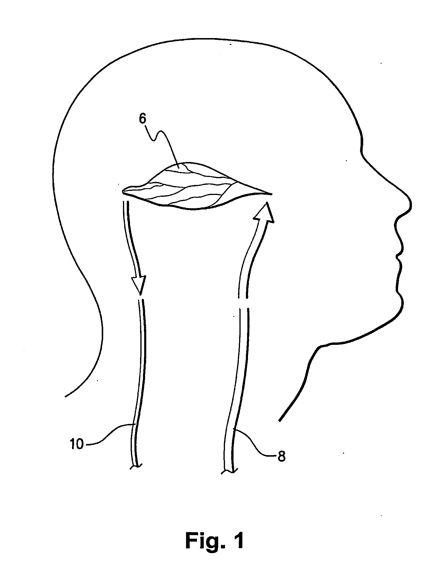
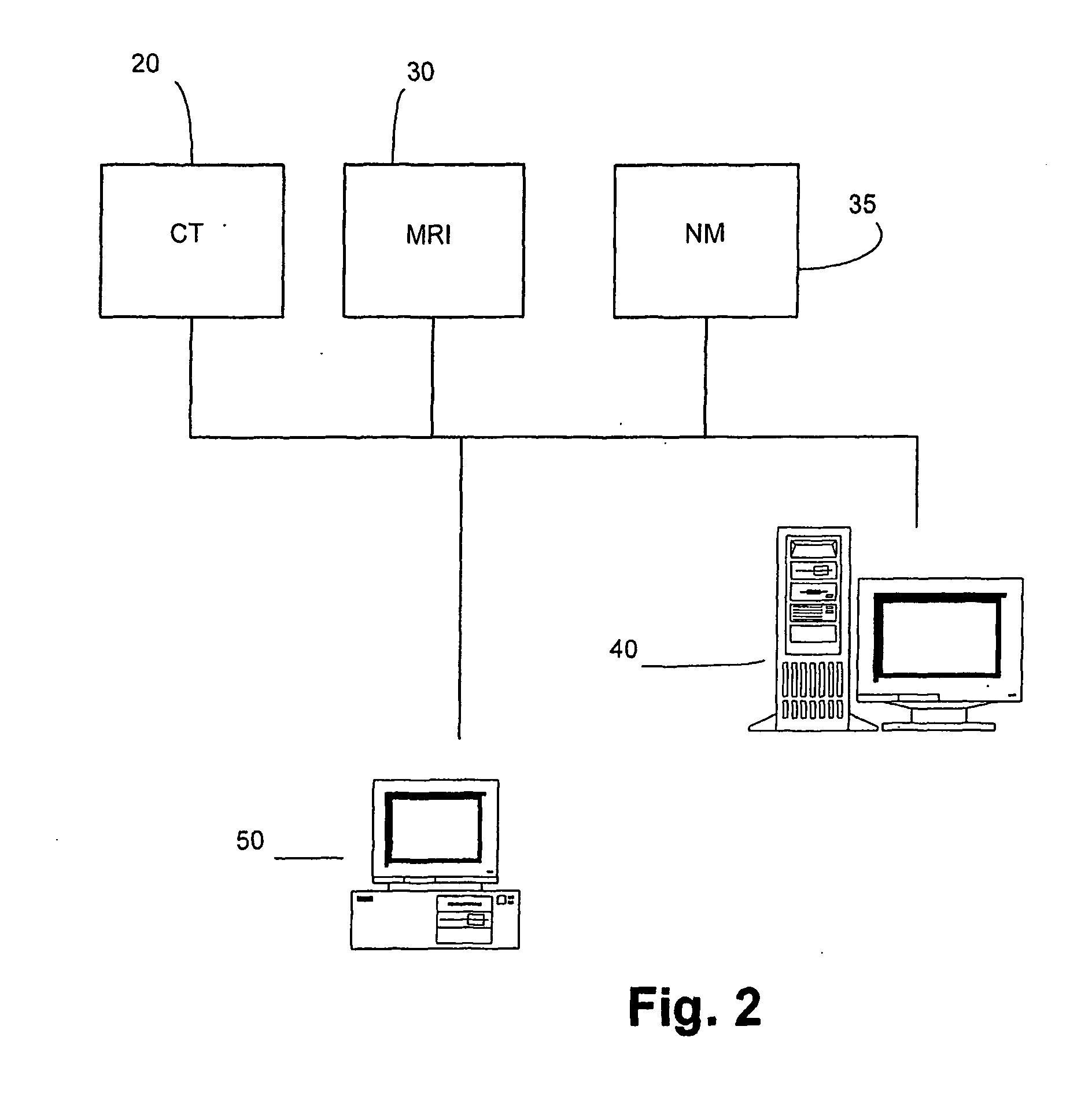
[0073] The present invention is particularly applicable to CT, MRI and MN imaging systems. A bolus of contrasting agents is introduced via a needle into a patient at, for example, the arm of the patient. However the bolus can be input to any other part of the patient. A region of interest (ROI) may be a tissue 6 in a part of the patient's brain as shown in FIG. 1. Alternatively, the ROI may be a pixel or a plurality of pixels, where many pixels represent a calculated image to produce one or more perfusion maps. Blood circulating throughout the patient will contain the contrast agent and in particular may be delivered to the tissue 6 via artery 8 and the blood flowing through the tissue 6 is returned to the heart via vein 10. Raw data and / or images collected by a scan, such as from a CT scanner 20, MRI scanner 30 or NM scanner 35 are forwarded to a data storage system 40 in the form of a Picture Archiving Communications System (PACS) in FIG. 2. A computer program operating on a proce...


PUM


Abstract

A method of deriving blood perfusion indices for a region of interest (ROI) of a subject, comprising the steps of administering a contrast agent to the subject during a dynamic imaging scan, converting signal intensity data from raw images of the scan into contrast agent concentration data, deriving parameters from the contrast agent concentration data using at least one transport function that accounts for delay and dispersion of the contrast agent, and calculating the blood perfusion indices from the derived parameters.

Description

FIELD OF THE INVENTION [0001] This invention relates to a method and system of obtaining improved data in blood perfusion measurements, and more particularly to a method and system of deriving blood perfusion indices for a region of interest of a subject. BACKGROUND TO THE INVENTION [0002] The process of measuring blood flow within a body of a subject non-invasively is useful in diagnosing and treating the subject. This is particularly the case where a part of a subject or patient, such as a tissue or organ, suffers from ischaemia due for example to a stroke. Determining perfusion indices including the blood flow through such a tissue or organ can provide important information to a physician in order to determine an appropriate treatment regime for the patient. [0003] A number of systems pertaining to blood flow information have been disclosed. In general, the systems involve a contrast agent delivered as an intravascular bolus during a dynamic imaging session such as computerized t...

Claims


Application Information

Patent Timeline
Patent Type & Authority Applications(United States)
IPC IPC(8): A61K49/00G06F19/00A61B5/026A61B5/0275A61B6/03G16B5/00G16B40/10
CPCA61B5/0263A61B5/0275A61B5/416A61B6/032A61B6/507A61B6/5217G06F17/10G06F17/13G06F19/24G06F19/12A61B6/481G06F17/11A61B5/4064G16B5/00G16B40/00G16H50/30G16B40/10
Inventor YANG, QING
Owner APOLLO MEDICAL IMAGING TECH
Why Eureka
Try Eureka