
Endovascular surgery device

a surgery device and endovascular technology, applied in the direction of intravenous devices, guide needles, infusion needles, etc., can solve the problems of affecting the healing time of patients, requiring at least five days of hospitalization, and the size of the catheter that can be used remains limited to the size of the femoral artery, so as to achieve the effect of advancing the catheter inside the artery all the way, and avoiding the need for hospitalization

Inactive Publication Date: 2006-04-20
FORMICHI MAXIME
View PDF12 Cites 18 Cited by
  • Summary
  • Abstract
  • Description
  • Claims
  • Application Information

AI Technical Summary

Benefits of technology

[0018] The object of the present invention is thus to provide a novel device enabling endovascular procedures to be performed via a laparoscopic approach.
[0019] More precisely, an object of the present invention is to provide a device enabling an artery to be punctured while controlling hemorrhaging and while inserting a guidewire into the artery laparoscopically, and subsequently making it possible to perform endovascular insertion of larger-sized catheters and endoprostheses via the laparoscopic approach.
[0020] To do this, the present invention provides a surgical device enabling a blood vessel to be punctured, in particular an artery, and a radiological guidewire to be inserted in endovascular manner, for use in laparoscopic or celioscopic endovascular surgery, in particular of the intra-abdominal blood vessels, the device comprising a first transparent flexible tube co-operating at its distal end with a hollow metal needle to which it is coupled, and in which it is possible to cause said radiological guidewire to pass, said first tube having, at its proximal end, closure means for closing said first tube, together with insertion means enabling a said radiological guidewire to be inserted in leaktight manner into said first tube.

Problems solved by technology

Firstly, the time required for healing requires at least five days of hospitalization when a surgical opening has been made.
Secondly, the size of catheters that can be used remains limited to the size of the femoral artery, i.e. about 7.2 mm to 8.4 mm (24 to 48 French).
Finally, some patients have iliac arteries suffering from stenoses, that are tortuous, and / or that are calcified, thus making it difficult or even impossible to advance the catheter inside the artery all the way to the site at which the endoprosthesis is to be delivered.
While the needle is inserted, bleeding occurs.
However, performing an endovascular procedure following a laparoscopic access, in particular to the intra-abdominal blood vessels, is not possible at present, for the following reasons.
Such hemorrhage naturally presents a danger to the patient.
In addition, it is not possible to suck out the blood since there will be a risk of simultaneously sucking out the gas previously inserted to enable the operation to be performed by the laparoscopic approach after inflating the abdomen.
Because blood absorbs light, this makes it difficult or even impossible to view the operation being performed, and thus makes it impossible in practice to perform an endovascular procedure under such conditions.

Method used


Image

  • Endovascular surgery device
  • Endovascular surgery device
  • Endovascular surgery device

Examples


Embodiment Construction

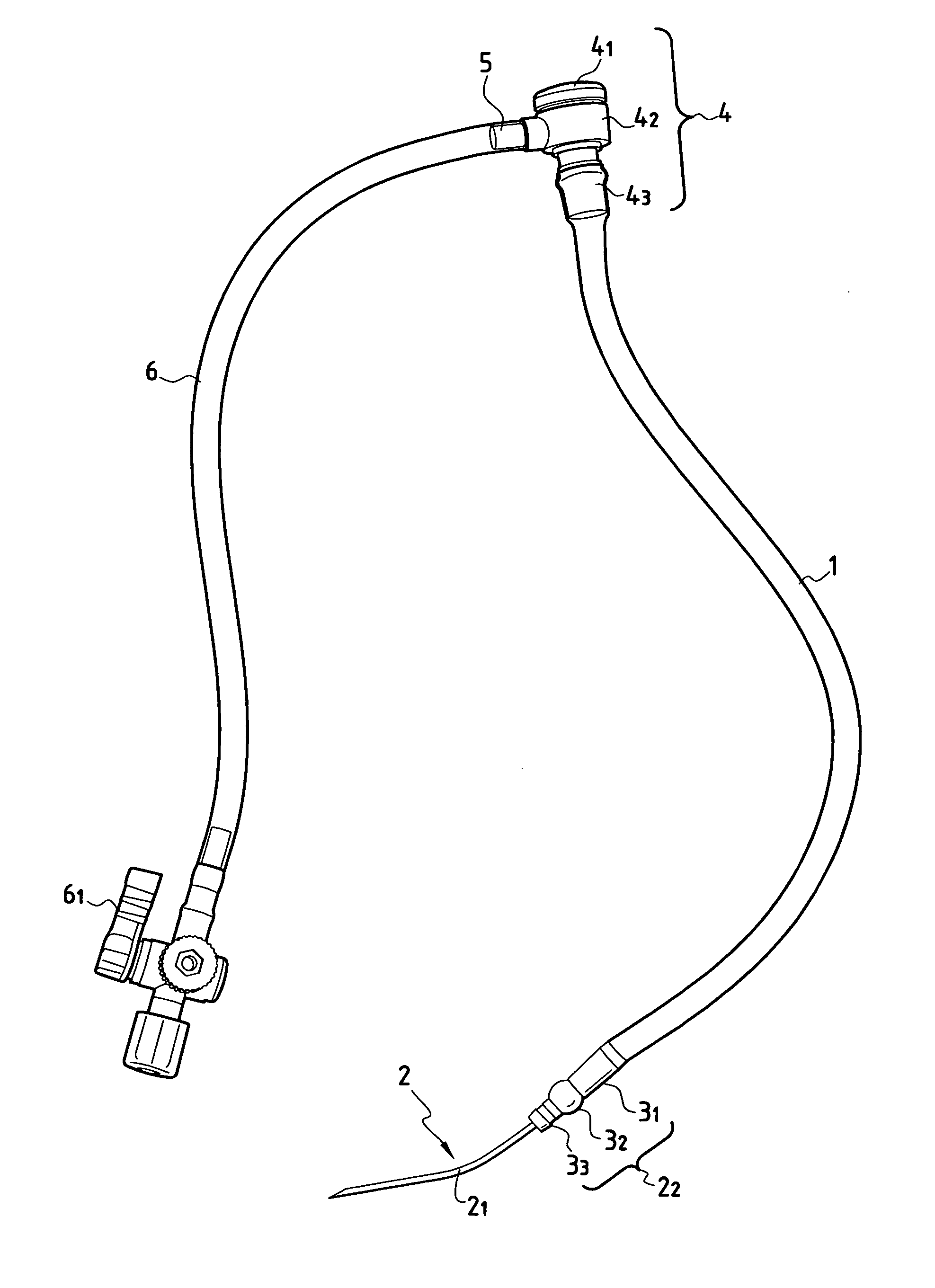
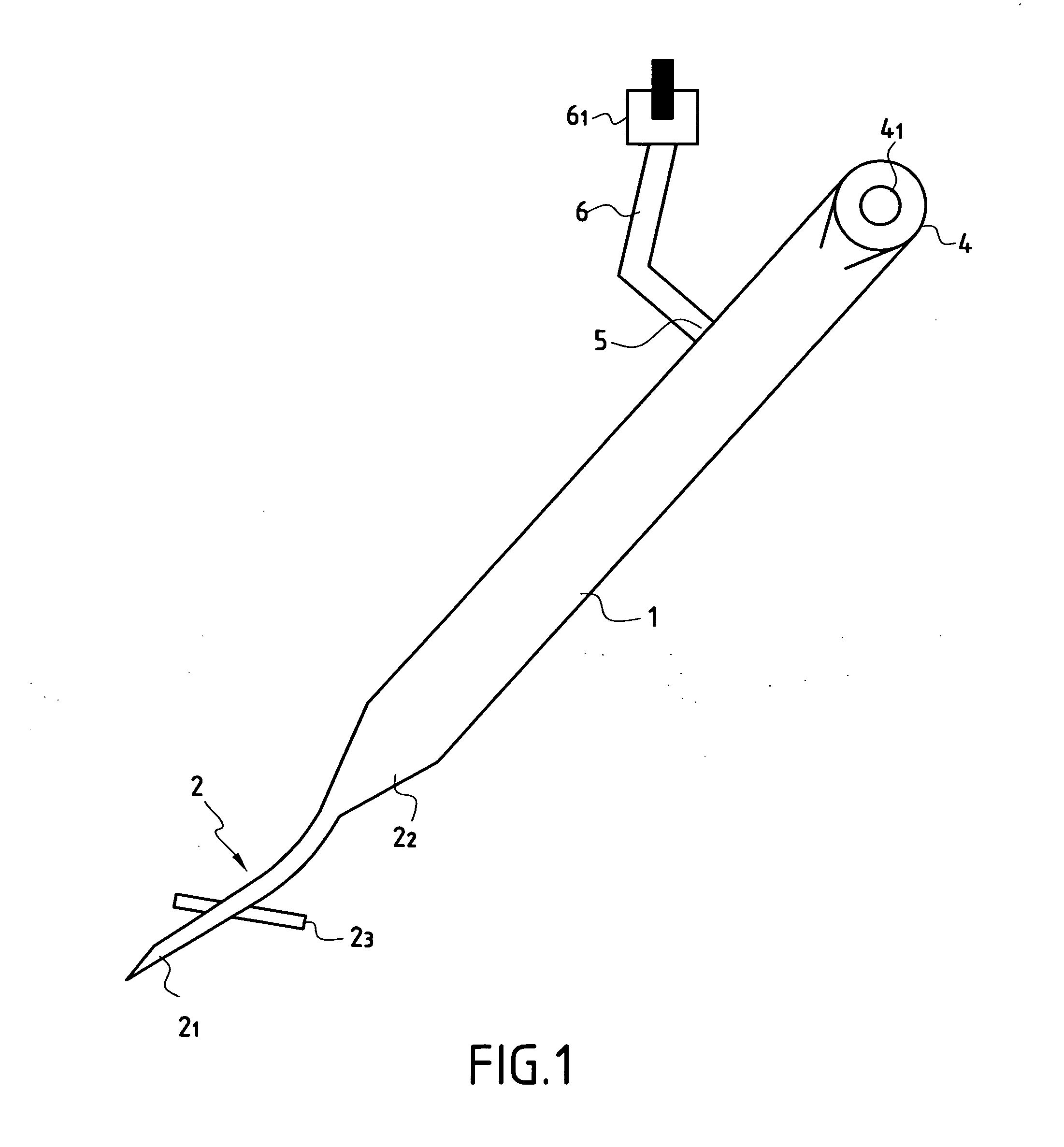
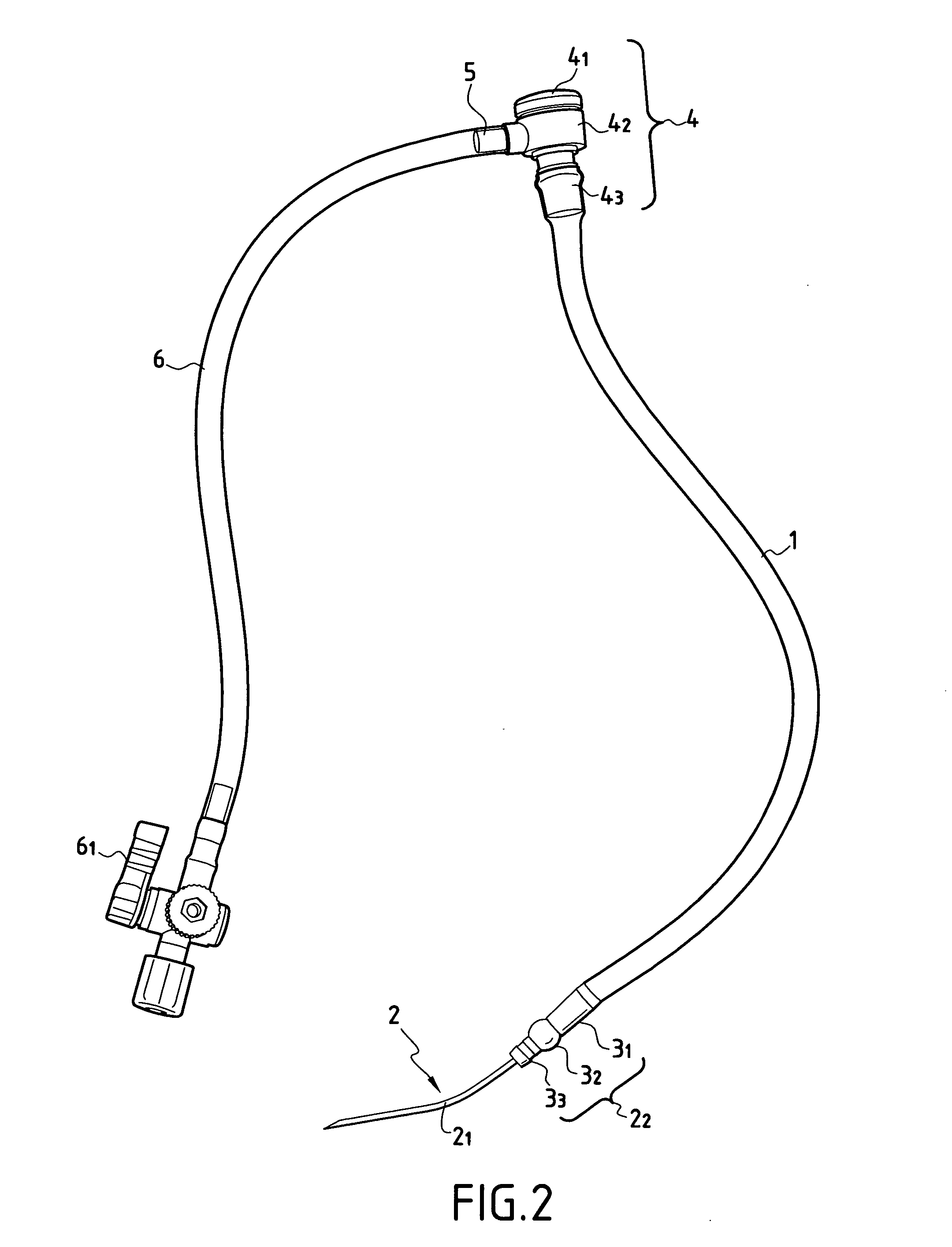
[0065] The device of the invention as shown in FIGS. 1 and 2 comprises: [0066] a said first transparent flexible tube 1 which, by way of illustration, presents a length of 20 cm to 50 cm and an outside diameter of 2 mm to 5 mm; and [0067] a hollow needle 2 comprising a curved hollow metal distal portion 21 with a first internal channel, and a proximal portion acting as a coupling element 22 for coupling with said first transparent tube 1, said coupling element having a second hollow internal channel.

[0068] During assembly, The distal end of said first tube 1, which is made of PVC, is engaged as a force-fit on the outside surface of a tubular sleeve 31 constituting the proximal portion of said coupling element 22.

[0069] The coupling element 22 has a hollow intermediate portion 32 constituted by an enlargement presenting an outline of rounded shape, having an outside diameter greater than the outside diameter of said first tubular sleeve 31. Said enlargement 32 is situated in line w...


PUM


Abstract

The present invention relates to surgical device enabling a blood vessel to be punctured, in particular an artery, and enabling a radiological guidewire to be inserted in endovascular manner for use in laparoscopic or celioscopic endovascular surgery, in particular of the intra-abdominal blood vessels, the device comprises a first transparent flexible tube co-operating at its distal end with a hollow metal needle to which it is coupled, and in which it is possible to cause a said radiological guidewire to pass, said first tube having, at its proximal end, closure means for closing said first tube, together with insertion means enabling a said radiological guidewire to be inserted in leaktight manner into said first tube.

Description

[0001] The present invention relates to a surgical device suitable for endovascular surgery, including interventional radiology. [0002] More particularly, the present invention relates to a device suitable for being implemented by a minimally invasive route, in particular by a laparoscopic and / or celioscopic route, in particular via the large intra-abdominal blood vessels. BACKGROUND OF THE INVENTION [0003] At present, endovascular surgery is performed for the following purposes and under the following conditions. [0004] The idea is to insert catheters, in particular balloon catheters and endoprostheses in order to treat arterial aneurysms or stenosis-forming lesions. [0005] These operations are performed either percutaneously, with various possible puncture sites, or by surgical approach via an artery, usually the femoral artery. [0006] Endoprostheses are inserted using catheters inserted inside the blood vessels. Percutaneous insertion can be performed for catheters of small size,...

Claims


Application Information

Patent Timeline
IPC IPC(8): A61M5/32A61B17/34A61M25/00A61M25/06A61M39/00
CPCA61B17/3415A61B17/3417A61B17/3478A61B17/3498A61M25/06A61M2025/0091
Inventor FORMICHI, MAXIME
Owner FORMICHI MAXIME
Learn More