Calibration of tissue densities in computerized tomography

a computerized tomography and tissue density technology, applied in the field of medical imaging using computerized tomography, can solve the problems of difficult reproducibility and more difficult measurement, and achieve the effect of reducing operator time, accurate and reproducible results, and being easy to apply

Inactive Publication Date: 2006-04-27
ARNOLD BEN A
View PDF77 Cites 32 Cited by
  • Summary
  • Abstract
  • Description
  • Claims
  • Application Information

AI Technical Summary

Benefits of technology

[0018] Unlike prior art devices in which image data was not calibrated, the disclosed invention presents a method to calibrate the image data such that a calibrated image detail edge can be quantitatively defined, located and relocated on follow up images. The reproducibility of physical measurements of target volumes, areas, and distances is greatly improved.
[0019] The embodiments of the present invention provide a method to calibrate the image to produce consistent and standardized results, independent of the measurement conditions discussed above (e.g., scanner type, beam energy, reconstruction software, patient size and composition, and the like). Clinical results will be more consistent, more accurate, and more precise. The ability to follow patients on follow-up exams to monitor disease progress will be greatly benefited.
[0020] The density differences in Hounsfield unit values between the soft tissues of the body are small, compared to bone density, and they are more difficult to measure. Microcalcifications in the vascular system of the body produce small density differences as well, and are difficult to measure reproducibly. The embodiments of the present invention overcome the small differences in the density of soft tissue or microcalcifications to facilitate measurements throughout the body, both in areas near the external phantom and in areas distant from the external phantom to overcome the inadequacies of the conventional methodology of QCT calibration with an external phantom. In particular, the embodiments of the present invention enable a method to quantify x-ray images that uses a hybrid calibration method that overcomes these limitations.

Problems solved by technology

The density differences in Hounsfield unit values between the soft tissues of the body are small, compared to bone density, and they are more difficult to measure.
Microcalcifications in the vascular system of the body produce small density differences as well, and are difficult to measure reproducibly.

Method used


Image

  • Calibration of tissue densities in computerized tomography
  • Calibration of tissue densities in computerized tomography
  • Calibration of tissue densities in computerized tomography

Examples


Embodiment Construction

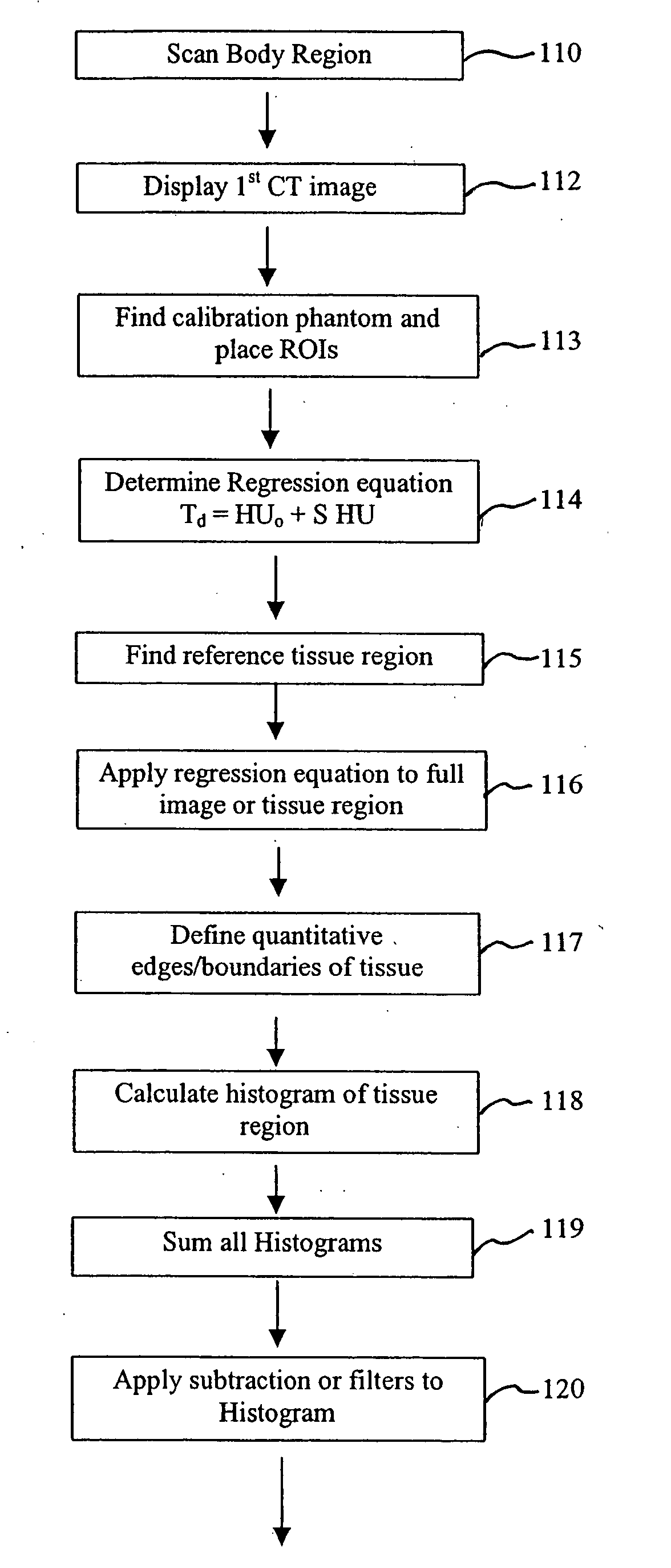
[0038] A preferred embodiment of the present invention is described below in connection with the flow chart of FIGS. 6A and 6B in view of the illustrations in FIGS. 1-5.

[0039] As illustrated by a step 110 in FIG. 6A, a CT scan of the subject is first taken with the subject lying on the reference calibration phantom. Several CT images are taken in a short time period. Preferably, CT images are taken using a multi-slice spiral scan CT scanner or using the EBCT Imatron scanner, although any CT scanner can be used for many tissues. Scans of the heart for coronary calcium analysis will require fast scan times to stop the motion of the beating heart.

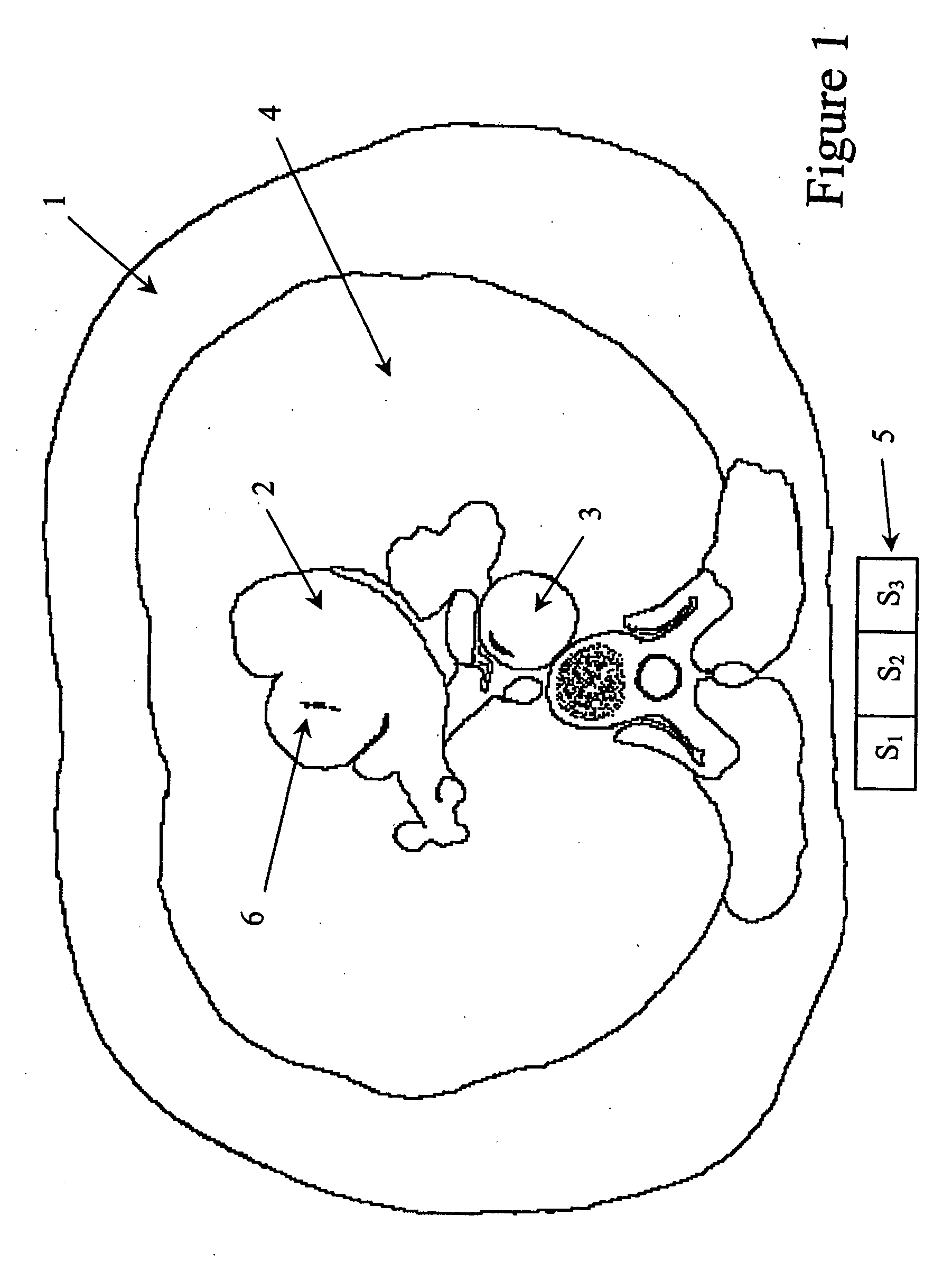
[0040] After reconstruction of the CT images in the scanner computer, a first cross-sectional image of the subject is displayed in a step 112 in FIG. 6A. FIG. 1 illustrates a representative depiction of such an image taken through the heart showing coronary calcium 6. The heart tissue 2, the aorta 3, the lung 4, the chest wall and a referenc...


PUM


Abstract

A hybrid calibration method uses a calibration phantom (exterior reference) scanned simultaneously with the patient, and one or more known tissues of the subject (interior reference) to create a hybrid calibration reference that improves the measurement of tissue densities throughout the body. In addition, the calibration method is used to quantitatively define boundaries of tissue and organs for more accurate measurements of lengths, areas and volumes. Another aspect of the invention uses the calibrated images to quantitatively preset absolute window / levels for filming and image display, which provides standardized viewing for diagnostic purposes.

Description

[0001] This application is a continuation of U.S. patent application Ser. No. 09 / 989,995, filed on Nov. 21, 2001, the entirety of which is hereby incorporated herein by reference.BACKGROUND OF THE INVENTION [0002] 1. Field of the Invention [0003] The present invention is in the field of medical imaging using computerized tomography (CT), and in particular, is directed to a method to determine tissue densities in the body of a subject and to provide calibrated display of images. [0004] 2. Description of the Related Art [0005] CT scanners have become a major diagnostic modality in modern medicine and are widely used for many types of exams. Most exams are performed by subjective viewing of the cross-sectional images on either film or electronic displays. This subjective viewing makes use of assumed quantitative image pixels, which define boundaries of tissues, organs, or foreign masses, and subjective discrimination of tissue types by density differences. Identification of diagnostic ...

Claims


Application Information

Patent Timeline
Patent Type & Authority Applications(United States)
IPC IPC(8): G06K9/00A61B6/00G06V10/147G06V10/25
CPCA61B5/02007A61B6/502A61B6/583G06K9/209G06K9/3233G06K2209/05G06T7/0012G06T2207/10081G06T2207/30004G06V10/147G06V10/25G06V2201/03
Inventor ARNOLD, BEN A.
Owner ARNOLD BEN A
Why Eureka
Try Eureka