

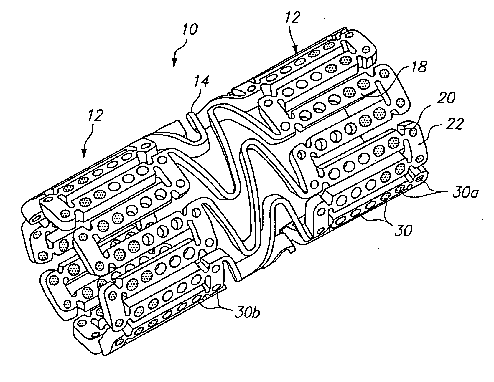
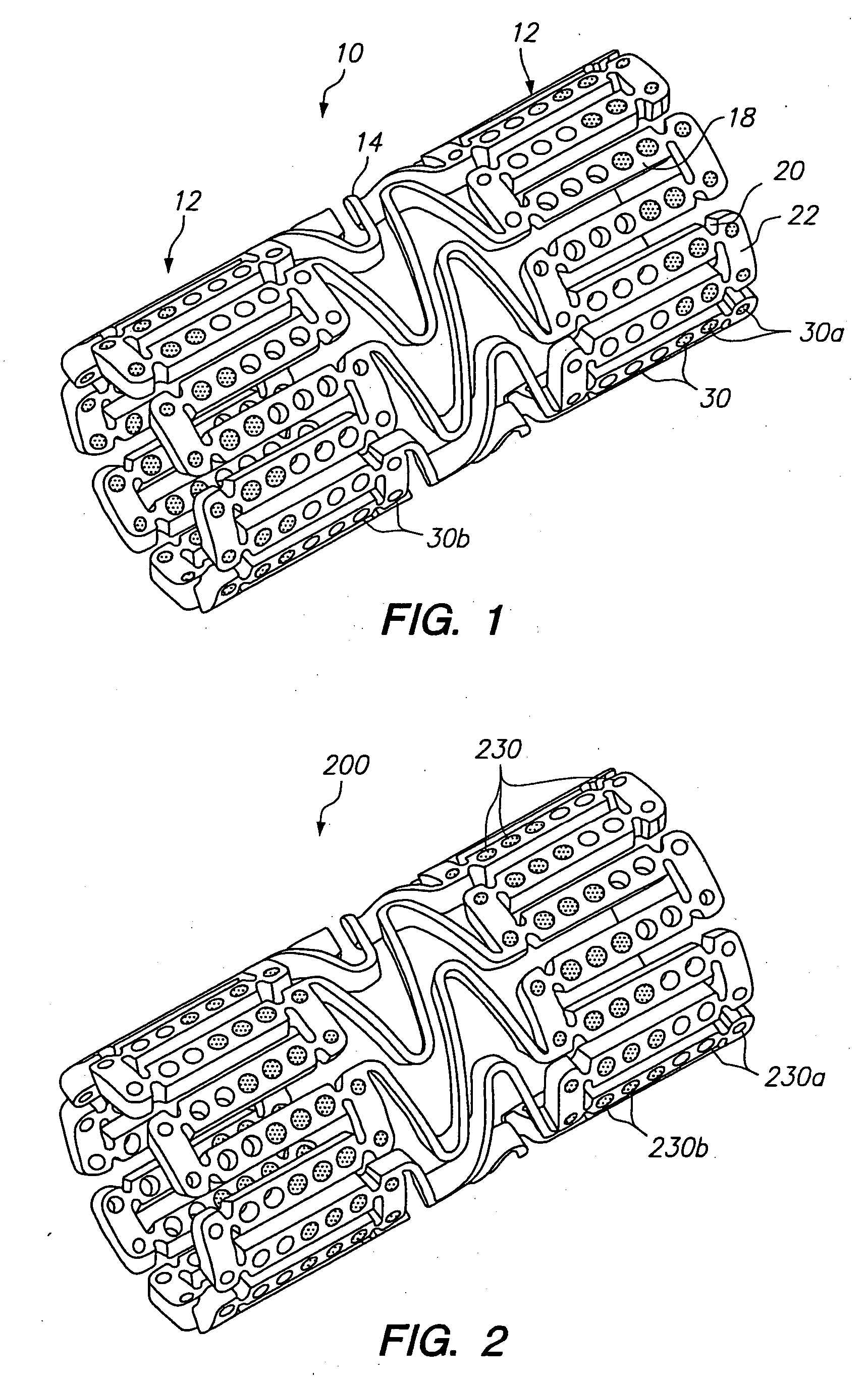
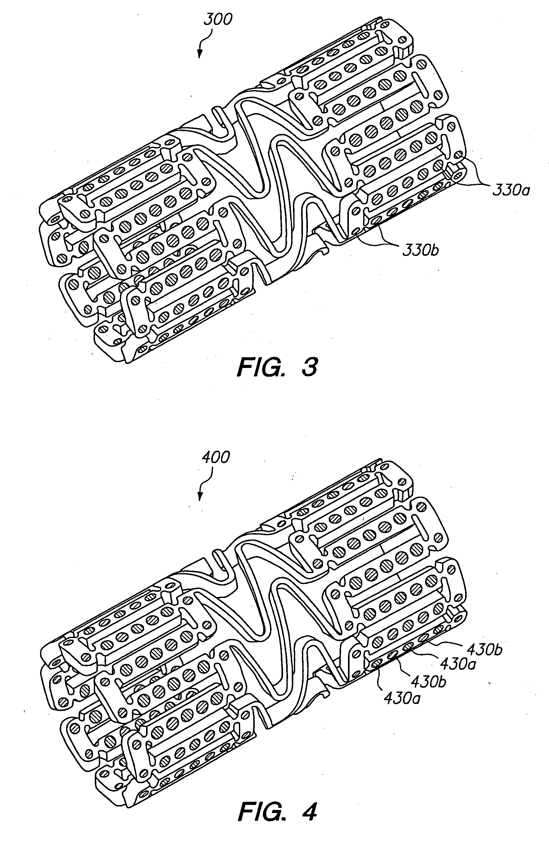
Expandable medical device with openings for delivery of multiple beneficial agents
a medical device and beneficial agent technology, applied in the field of tissue-supporting medical devices, can solve the problems of increasing trauma and risk to patients, reducing the mechanical expansion properties of the stent, and increasing the volume of beneficial agents, so as to improve adversely affecting the mechanical expansion properties of the stent, and increasing the effective wall thickness of the sten
- Summary
- Abstract
- Description
- Claims
- Application Information
AI Technical Summary
Benefits of technology
Problems solved by technology
Method used
Image

Examples
example 1
[0071]FIG. 7 illustrates a dual drug stent 700 having an anti-inflammatory agent and an antiproliferative agent delivered from different holes in the stent to provide independent release kinetics of the two drugs which are specifically programmed to match the biological processes of restenosis. According to this example, the dual drug stent includes the anti-inflammatory agent pimecrolimus in a first set of openings 710 in combination with the antiproliferative agent paclitaxel in a second set of openings 720. Each agent is provided in a matrix material within the holes of the stent in a specific inlay arrangement designed to achieve the release kinetics shown in FIG. 8. Each of the drugs are delivered primarily murally for treatment of restenosis.
[0072] As shown in FIG. 7, pimecrolimus is provided in the stent for directional delivery to the mural side of the stent by the use of a barrier 712 at the luminal side of the hole. The barrier 712 is formed by a biodegradable polymer. Th...
example 2
[0077] According to this example, the dual drug stent includes the Gleevec in the first set of openings 710 in combination with the antiproliferative agent paclitaxel in the second set of openings 720. Each agent is provided in a matrix material within the holes of the stent in a specific inlay arrangement designed to achieve the release kinetics shown in FIG. 8.
[0078] The Gleevec is delivered with a two phase release including a high initial release in the first day and then a slow release for 1-2 weeks. The first phase of the Gleevec release delivers about 50% of the loaded drug in about the first 24 hours. The second phase of the release delivers the remaining 50% over about one-two weeks. The paclitaxel is loaded within the openings 720 in a manner which creates a release kinetic having a substantially linear release after the first approximately 24 hours, as shown in FIG. 8 and as described above in Example 1.
[0079] The amount of the drugs delivered varies depending on the si...
example 3
[0080] According to this example, the dual drug stent includes the PKC-412 (a cell growth regulator) in the first set of openings in combination with the antiproliferative agent paclitaxel in the second set of openings. Each agent is provided in a matrix material within the holes of the stent in a specific inlay arrangement designed to achieve the release kinetics discussed below.
[0081] The PKC-412 is delivered at a substantially constant release rate after the first approximately 24 hours, with the release over a period of about 4-16 weeks, preferably about 6-12 weeks. The paclitaxel is loaded within the openings in a manner which creates a release kinetic having a substantially linear release after the first approximately 24 hours, with the release over a period of about 4-16 weeks, preferably about 6-12 weeks.
[0082] The amount of the drugs delivered varies depending on the size of the stent. For a 3 mm by 6 mm stent the amount of PKC-412 is about 100 to about 400 μg, preferably...
PUM
| Property | Measurement | Unit |
|---|---|---|
| Fraction | aaaaa | aaaaa |
| Fraction | aaaaa | aaaaa |
| Time | aaaaa | aaaaa |
Abstract
Description
Claims
Application Information