Apparatus and method for immunological labeling for thin tissue sections

a technology of immunological labeling and apparatus, applied in the direction of biochemistry apparatus, biochemistry apparatus and processes, preparing samples for investigation, etc., can solve the problems of high labor costs, large number of samples to be processed simultaneously, and constant attention to the process of the operator

Inactive Publication Date: 2006-06-15
LEICA MICROSYSTEMS GMBH
View PDF2 Cites 6 Cited by
  • Summary
  • Abstract
  • Description
  • Claims
  • Application Information

AI Technical Summary

Benefits of technology

[0012] It is therefore the object of the invention to make available an apparatus for immune labeling of thin tissue sections which effectively permits multiple thin tissue sections to be processed simultaneously with reproducible quality, fully automatically and according to an individually definable protocol, and with no intervention by the operator.
[0014] A further object of the invention is to make available a method for immune labeling of thin tissue sections which effectively permits multiple thin tissue sections to be processed simultaneously with reproducible quality, fully automatically and according to an individually definable protocol, and with no intervention by the operator.
[0023] The apparatus according to the present invention is particularly advantageous because the apparatus is equipped with a carrier plate on which the thin tissue sections are affixed in a defined pattern. Also provided is at least one slide on which the treatment liquid is applied in the form of liquid droplets, the arrangement of the liquid droplets corresponding to the arrangement of the thin tissue sections on the carrier plate. Each of the slides that is equipped with liquid droplets is introduced into a transport container. The transport container is embodied in the form of a trough, the trough comprising a peripheral delimiting wall that is closed off by a base. The base (50) of one transport container is the cover of a transport container arranged beneath it. The transport containers are stacked in a first and a second station (20, 21). Stacking of the transport containers is particularly advantageous because evaporation of the liquid droplets applied on the slide is thereby prevented. To ensure a certain moisture level in the enclosed space around the slides, a moisture-emitting medium is placed in the transport container.
[0025] Multiple metal grids are mounted on the carrier plate, so that the number of samples or thin tissue sections to be processed simultaneously is greatly increased. For treatment of the thin tissue sections, the slides provided in transport containers are brought successively, in accordance with the procedure required for the individual treatment process, into defined contact with the carrier plate so that the thin tissue section is wetted with the treatment liquid. The purpose here is to move the requisite slide into the treatment position with respect to the carrier plate.

Problems solved by technology

At present, these washing operations are performed manually, in a laborious process in which individual droplets of the aqueous buffer solution or labeling solution are applied with a pipette onto a hydrophobic substrate (e.g. Parafilm®, Parlodion®, Colloidion, or Formfan®).
It is readily apparent that this manual process requires constant attention by the operator, and entails high labor costs because of the large time requirement.
The number of samples to be processed simultaneously is greatly limited, and errors by the operator while precisely pipetting and positioning liquid droplets with very small volumes cannot be excluded.
In addition, evaporation of the liquid droplets during longer-duration standard protocols constitutes a major problem.
This apparatus is not suitable for the implementation of immunological labeling techniques themselves, since the antibody-containing labeling solutions that are used are very expensive and are therefore employed in only the smallest possible volumes.

Method used


Image

  • Apparatus and method for immunological labeling for thin tissue sections
  • Apparatus and method for immunological labeling for thin tissue sections
  • Apparatus and method for immunological labeling for thin tissue sections

Examples


Embodiment Construction

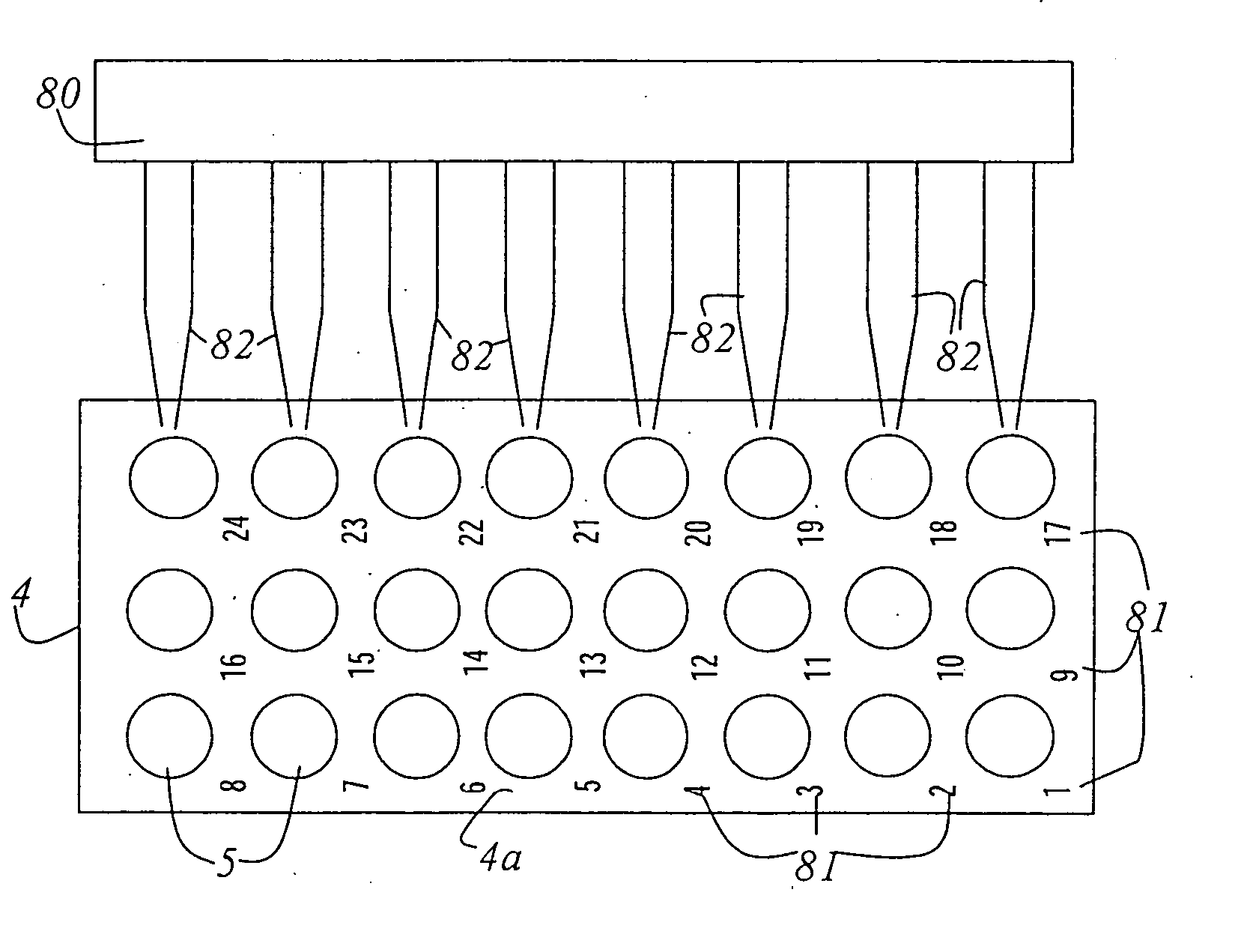
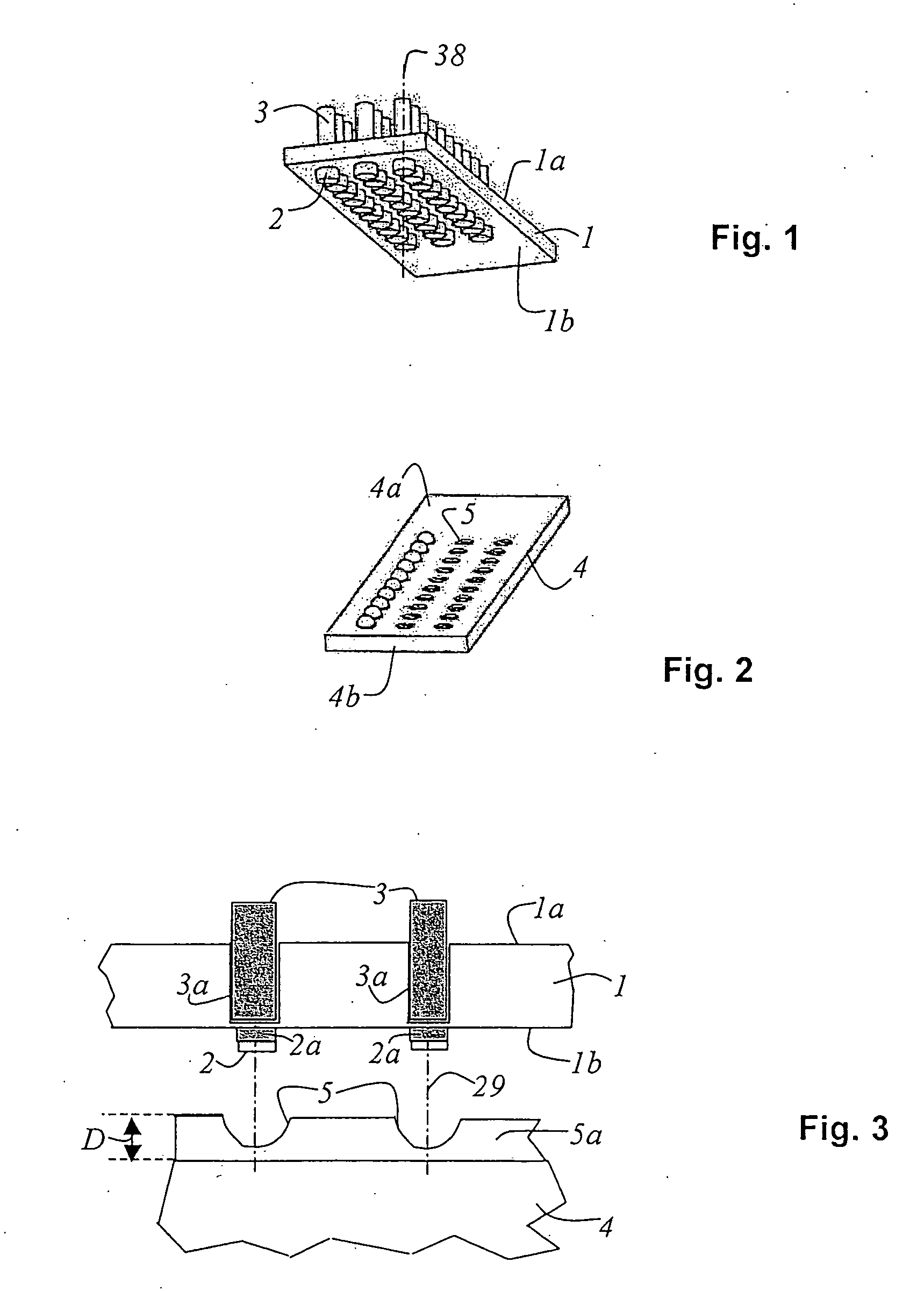
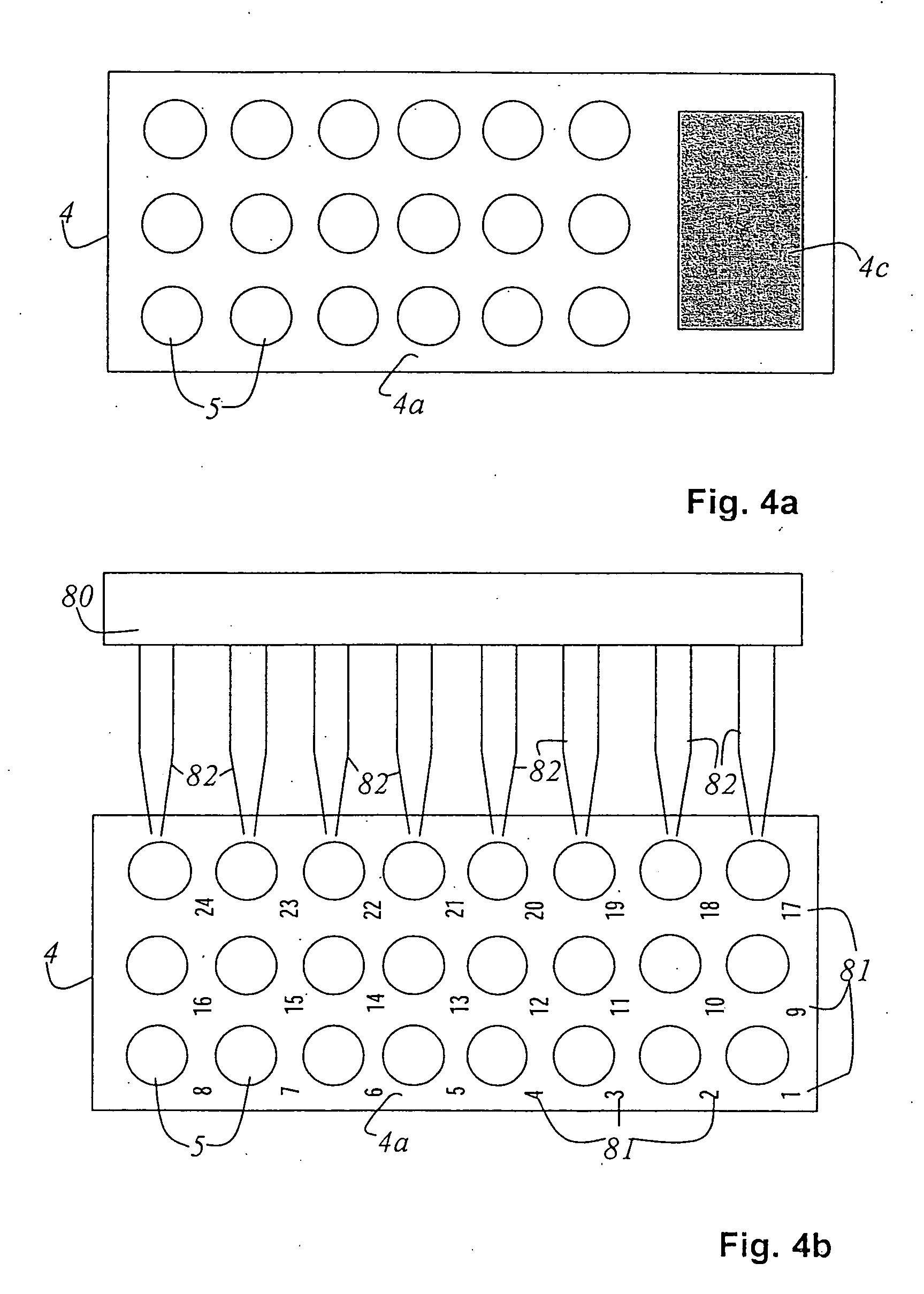
[0044]FIG. 1 shows a carrier plate 1 that defines an upper side 1a and a lower side 1b. On lower side 1b, carrier plate 1 possesses marked positions at which metal grids 2 having thin tissue sections 2a (see FIG. 3) are positioned. The positions of metal grids 2 on carrier plate 1 are elevated. This prevents liquid bridges from forming between the individual metal grids 2 upon contact with the liquid droplets. Located on upper side 1a of carrier plate 1, opposite the positions of metal grids 2, are orifices 3a (see FIG. 3) which contain magnets 3, for example permanent magnets. They cause metal grids 2 to be held in place on lower side 1b of carrier plate 1 by magnetic force. The spacing between metal grid 2 and magnet 3 should be kept as small as possible (1 is preferably made of a dimensionally stable nonmagnetic material, preferably aluminum, brass, or fiber-reinforced plastics, and advantageously is hydrophobically coated (e.g. with a Teflon pressure coating) on lower side 1b th...


PUM

PropertyMeasurementUnit
residence timeaaaaaaaaaa
diameteraaaaaaaaaa
sizeaaaaaaaaaa

Abstract

An apparatus and a method for immunological labeling of thin tissue sections are disclosed. The treatment fluid is applied in the form of liquid droplets onto at least one slide, the arrangement of liquid droplets corresponding to the arrangement of the thin tissue sections on the carrier plate. Each slide is retained in a transport container, the transport container being embodied in the form of a trough which comprises a peripheral delimiting wall that is closed off by a base. The base of one transport containers constitutes the cover of a transport container arranged beneath it. The transport containers are stacked in a first and a second station.

Description

CROSS REFERENCE TO RELATED APPLICATIONS [0001] This application is a divisional application of U.S. patent application Ser. No. 10 / 789,529, filed Feb. 27, 2004. U.S. patent application Ser. No. 10 / 789,529 claims priority of the German patent application 103 09 211.0, filed Feb. 28, 2003, which is incorporated by reference herein.FIELD OF THE INVENTION [0002] The present invention concerns an apparatus for immunological labeling for thin tissue sections. In particular, the invention concerns an apparatus for immunological labeling of thin tissue sections having a carrier plate on which the thin tissue sections are affixed in a defined pattern, and having at least one slide on which the treatment liquid is applied in the form of liquid droplets, the arrangement of the liquid droplets corresponding to the arrangement of the thin tissue sections on the carrier plate. [0003] Furthermore, the present invention concerns a method for immunological labeling for thin tissue sections. BACKGROU...

Claims


Application Information

Patent Timeline
Patent Type & Authority Applications(United States)
IPC IPC(8): G01N33/00G01N33/48B01L9/00C12M1/34G01N1/28G01N1/30G01N1/31G01N33/53G01N33/543G01N35/04
CPCB01L9/52G01N1/30G01N1/312Y10T436/13G01N33/54366G01N35/04G01N33/543
Inventor PLANK, HEINZ
Owner LEICA MICROSYSTEMS GMBH
Why Eureka
Try Eureka