

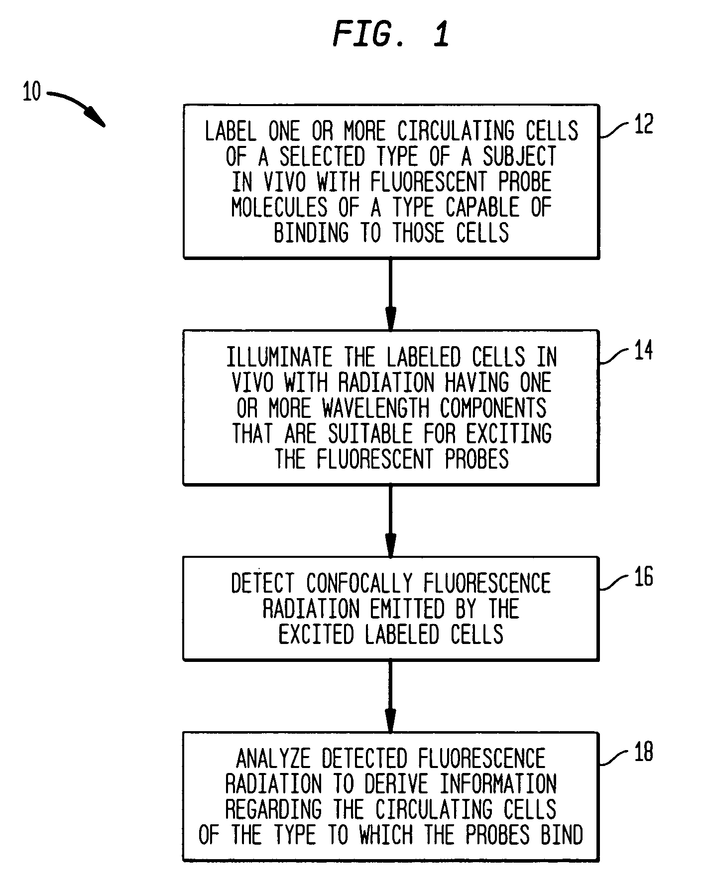
In vivo flow cytometry system and method
a flow cytometry and flow cytometry technology, applied in the field of flow cytometry systems and methods, can solve the problems of inability to accurately track cells flowing at a high speed, and inability to accurately measure cells, etc., to achieve the effect of minimizing interferen
- Summary
- Abstract
- Description
- Claims
- Application Information
AI Technical Summary
Benefits of technology
Problems solved by technology
Method used
Image

Examples
example
[0065] A number of measurements were performed in mice to demonstrate the efficacy of the methods and systems according to the teachings of the invention for real-time in vivo quantification of circulating cells in a subject. For these measurements, human red blood cells (RBC) were isolated and labeled ex vivo with 0.1 mM DiD (Molecular Probe V22887) (a lipophilic dye that binds to cell membranes). The labeled cells were then injected into a mouse's circulation through the tail vein. In these illustrative measurements, an ex vivo labeling procedure, rather than direct injection into the mouse's circulatory system, was employed so as to limit the fraction of labeled RBC's to less than about 1% in order to avoid overlap of signal pulses.
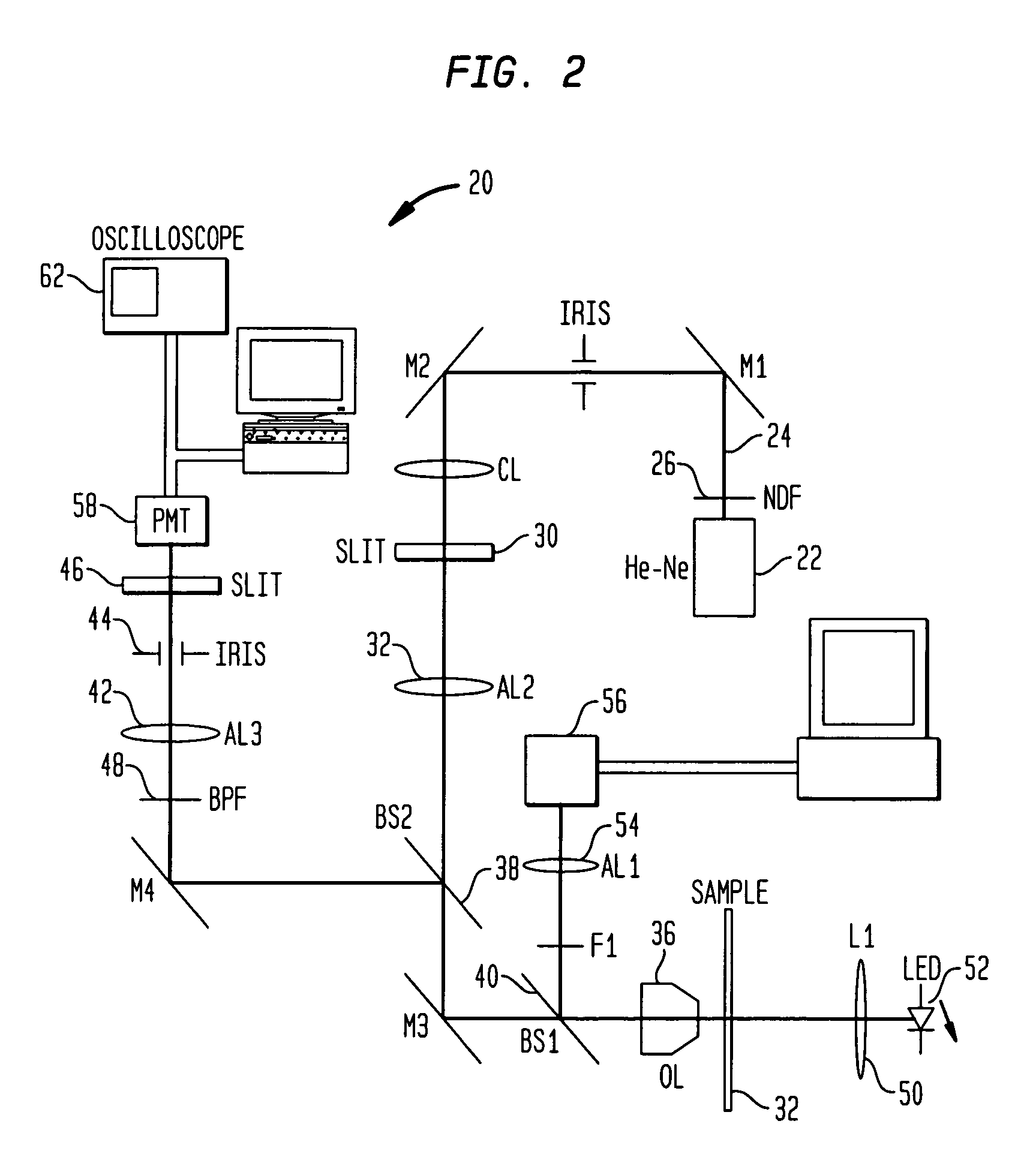
[0066] An instrument such as the above system 20 was employed to acquire the fluorescent data. More specifically, a He—Ne light source was employed for generating excitation radiation. To identify a blood vessel location for measurement, the mouse's e...
PUM
| Property | Measurement | Unit |
|---|---|---|
| Wavelength | aaaaa | aaaaa |
| Wavelength | aaaaa | aaaaa |
| Time | aaaaa | aaaaa |
Abstract
Description
Claims
Application Information