Methods for staining cells for identification and sorting

a cell and staining technology, applied in nanoinformatics, instruments, veterinary instruments, etc., can solve the problems of complex cells that outweigh the advantages, fixation procedures often incur artifacts, and damage to the rest of the cell structure, so as to facilitate the introduction of dye and enhance sperm viability

Inactive Publication Date: 2006-08-03
ARRYX INC
View PDF4 Cites 10 Cited by
  • Summary
  • Abstract
  • Description
  • Claims
  • Application Information

AI Technical Summary

Benefits of technology

[0016] In a first aspect, the invention discloses methods for staining cells, such as sperm, including bovine sperm, wherein the sperm are mixed with a dye of choice and then electroporating them to facilitate the introduction of the dye. The sperm can be incubated at temperatures that enhance sperm viability, typically equal to or less than 39° C.

Problems solved by technology

These techniques suffer from the preparation processes that allow for the visualization.
Fixation procedures often incur artifacts; for example, in the early days of electronic microscopy (EM), multilamellar bodies were observed but were later understood to be mostly by-products of the fixation protocols, not actual structures found in living mammalian cells.
While fixation protocols do preserve some of the cell structure, there are many structures that are difficult to preserve, or when preserved under appropriate conditions, the rest of the cell architecture is destroyed.
This approach does offer the advantages of non-fixed cells; however, the time and expense to purify a target polypeptide, conjugate it to a dye, and then to microinject (a task requiring specialized equipment, experience, skill and patience) the complex into a cell often outweigh the advantages.
Furthermore, only limited numbers of cells could be examined at any given time due to the limitations of microinjection.
While this approach is far superior to previous methods, many extra, time-consuming, steps are required from identifying the protein of interest to actually visualizing it in a living cell.
In each case, however, the challenge of introducing the dye or stain into a living cells to the appropriate target area is hindered by the cell membrane which provides a barrier to cells from the outside world.
Available procedures are few and when available, often face uncompromising challenges.

Method used


Image

  • Methods for staining cells for identification and sorting
  • Methods for staining cells for identification and sorting

Examples


example 1

In vivo Staining of Sperm Cells with a DNA-Specific Dye

[0082] Methods and Materials

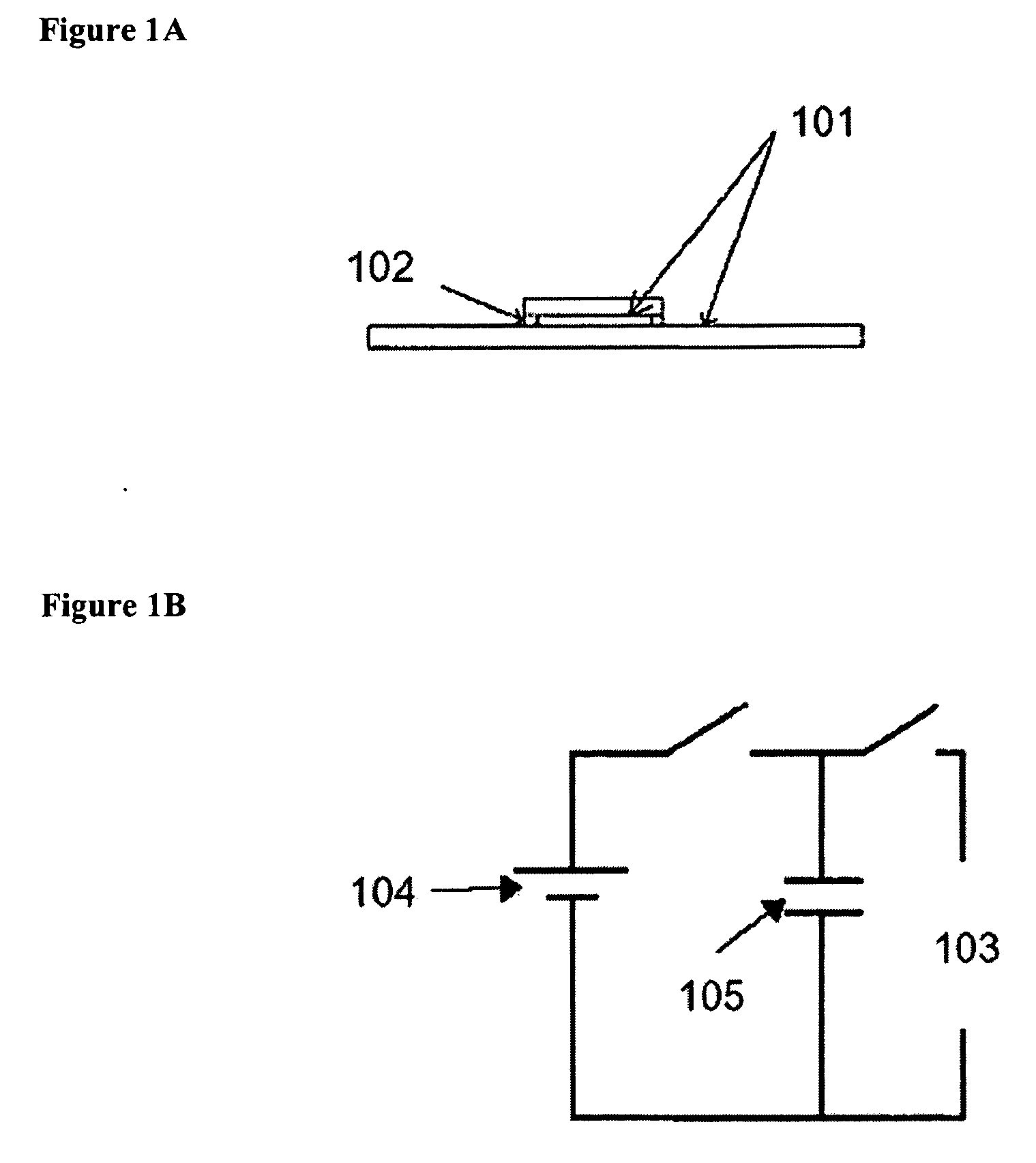
[0083] Electroporation unit A sample cell was formed by two parallel glass slides coated with 1,500-2000 angstroms (Å) of indium tin oxide (ITO). The slides were separated by fragments of number zero glass cover slips, yielding a slide separation of 100 millimeters (mm).

[0084] The sample cell was connected to a resistor-capacitator (RC) circuit by alligator clips. A direct current (DC) power supply was used to charge a capacitor. When a switch was thrown, the discharging capacitor generated a time-dependant and spatially uniform electric field across the sample. An oscilloscope was used to monitor the voltage across the sample cell as a function of time.

[0085] The RC circuit formed by the sample cell and capacitor allowed for a well-controlled electric field to be generated. The resistance (R) of the circuit was left floating—that is, determined by the geometry and content of the sample cell. Typi...


PUM

PropertyMeasurementUnit
temperatureaaaaaaaaaa

Abstract

The present invention provides novel methods of cell staining, such as bovine sperm, using electroporation or osmolality treatments at viability-enhancing temperatures. Furthermore, methods of highly efficient cell sorting that are especially suitable in sorting bovine sperm using novel cell staining procedures are also provided.

Description

CROSS-REFERENCE TO RELATED APPLICATIONS [0001] This application incorporates by reference the related application, METHOD AND APPARATUS FOR SORTING CELLS, Ser. No. ______, filed ______, and Representative Docket Number 089000-0138.STATEMENT REGARDING FEDERALLY SPONSORED RESEARCH OR DEVELOPMENT [0002] Not Applicable. INCORPORATION-BY-REFERENCE OF MATERIAL SUBMITTED ON A COMPACT DISC [0003] Not Applicable. FIELD OF THE INVENTION [0004] The invention relates to techniques and systems for visualizing live cells using novel staining procedures. BACKGROUND OF THE INVENTION [0005] The in vivo identification of a target cell population is required, and required quickly, in many industries. Such applications include those where the selected cells are destined for other applications that require the cells to be living after identification. For example, cells are processed using fluorescence-activated cell sorting, where cells are cultured and expanded in vitro after sorting, or in sperm sorti...

Claims


Application Information

Patent Timeline
Patent Type & Authority Applications(United States)
IPC IPC(8): C12Q1/68G01N33/48
CPCB82Y5/00B82Y10/00G01N33/5005G01N33/689G01N15/1425G01N21/6428G01N2015/1081G01N2021/6439A61D19/04C12N5/061C12N5/0612G01N1/30G01N15/14G01N2015/0065G01N2015/1006G01N2015/149
Inventor ANDERSON, AMY L.KNUTSON, CHRISTOPHER R.MUETH, DANIELPLEWA, JOSEPHTANNER, EVAN
Owner ARRYX INC
Why Eureka
Try Eureka