Method and system for automatically segmenting organs from three dimensional computed tomography images

a computed tomography and organ technology, applied in image analysis, image enhancement, instruments, etc., can solve the problems of time-consuming and laborious, manual tracing of outlines on a contiguous set of 2d slices and then combining them, and in some cases, impossibl

Inactive Publication Date: 2006-10-12
GENERAL ELECTRIC CO
View PDF16 Cites 57 Cited by
  • Summary
  • Abstract
  • Description
  • Claims
  • Application Information

AI Technical Summary

Problems solved by technology

Also, the patient may have some type of individual differences or abnormalities regarding even healthy tissues.
Manually tracing the outlines on a contiguous set of 2D slices and then combining them can be time consuming and labor intensive.
A very small intensity difference causes separation of each organ, if “touching” slice images are contained within the region of interest (ROI), to be very difficult and, in some cases, impossible.
Additionally, between acquisitions of the slice images, abdominal organs are displaced by respiratory motion, resulting in significantly jagged outlines in planes tilted relative to a plane of the slice image.

Method used


Image

  • Method and system for automatically segmenting organs from three dimensional computed tomography images
  • Method and system for automatically segmenting organs from three dimensional computed tomography images
  • Method and system for automatically segmenting organs from three dimensional computed tomography images

Examples


Embodiment Construction

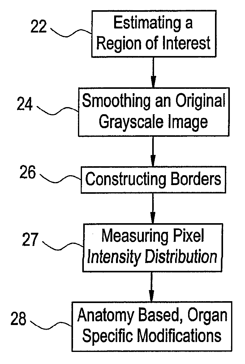
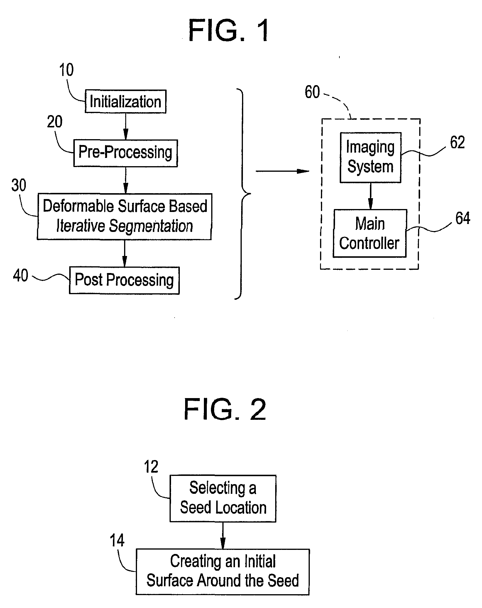
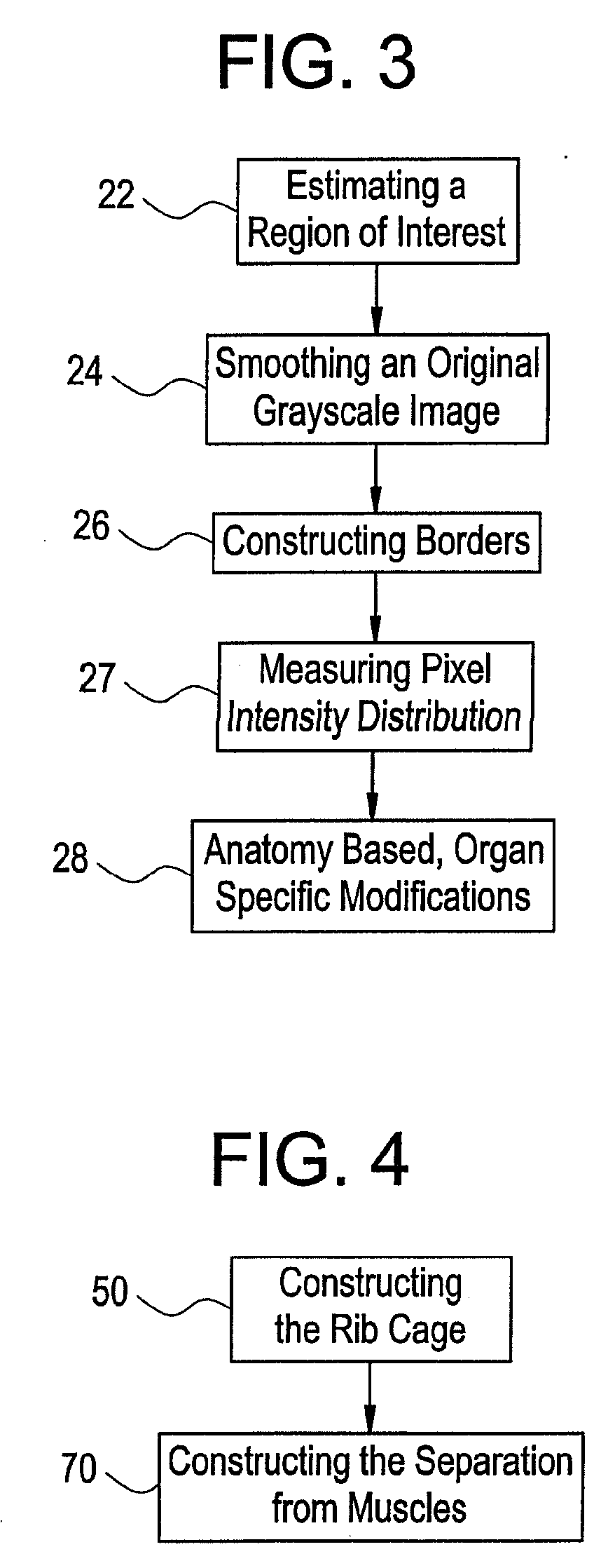
[0027] Although a detailed description of a method and system for automatically segmenting abdominal organs from three dimensional (3D) images follows, it will be appreciated by one skilled in the art that the method and system that follow are also suitable for segmenting other organs, especially organs located in a soft tissue environment.

[0028] An embodiment of the invention may be embodied in the form of computer-implemented processes and apparatuses for practicing those processes. The present invention may also be embodied in the form of computer program code containing instructions embodied in tangible media, such as floppy diskettes, CD-ROMs, hard drives, or any other computer readable storage medium, wherein, when the computer program code is loaded into and executed by a computer, the computer becomes an apparatus for practicing the invention. The present invention may also be embodied in the form of computer program code, for example, whether stored in a storage medium, lo...


PUM


Abstract

A method for automatically segmenting image data of an object includes selecting a seed, creating an initial surface around the seed, preprocessing the image data, running a deformable surface algorithm on the image data and post processing the image data. Selecting the seed includes at least one of selecting one seed point within the object, selecting two seed points within the object with one seed point being selected at an upper end of the object and the other seed point being selected at a lower end of the object if the object is a first organ, and selecting a curved segment within the object if the object is a second organ.

Description

BACKGROUND OF THE INVENTION [0001] The present invention relates generally to image systems and, more particularly, to systems and methods for segmenting an organ in a plurality of images. [0002] During clinical diagnosis, a patient's internal anatomy is imaged to determine how a disease has progressed. An infected tissue (such as tumor mass) shows some differences from a normal tissue. Also, the patient may have some type of individual differences or abnormalities regarding even healthy tissues. [0003] Several modalities are used to generate images of the patient's internal anatomy or functionality, suitable for diagnostic purposes, radiotherapy treatment, or for surgical planning. Exemplary modalities include conventional X-ray plane film radiography; computed tomography (“CT”) imaging, magnetic resonance imaging (“MRI”); and nuclear medicine imaging techniques, such as positron emission tomography (“PET”) and single photon emission computed tomography (“SPECT”). [0004] In a case ...

Claims


Application Information

Patent Timeline
Patent Type & Authority Applications(United States)
IPC IPC(8): G06K9/00
CPCG06T7/0083G06T2207/30084G06T2207/20101G06T7/0089G06T7/12G06T7/149
Inventor FIDRICH, MARTAMATE, EORSNYUL, LASZLO G.KUBA, ATTILAKISS, BENCE
Owner GENERAL ELECTRIC CO
Why Eureka
Try Eureka