Dedicated display for processing and analyzing multi-modality cardiac data

a multi-modality, cardiac data technology, applied in the field of medical imaging, can solve the problems of not allowing users to view fused volumes in 3d space, operating with modeled hearts, and not actual heart images

Inactive Publication Date: 2006-10-26
SIEMENS MEDICAL SOLUTIONS USA INC
View PDF6 Cites 91 Cited by
  • Summary
  • Abstract
  • Description
  • Claims
  • Application Information

AI Technical Summary

Problems solved by technology

Although proven to be quite useful, this technique does not allow users to view fused volumes in 3D spa

Method used


Image

  • Dedicated display for processing and analyzing multi-modality cardiac data
  • Dedicated display for processing and analyzing multi-modality cardiac data
  • Dedicated display for processing and analyzing multi-modality cardiac data

Examples


Embodiment Construction

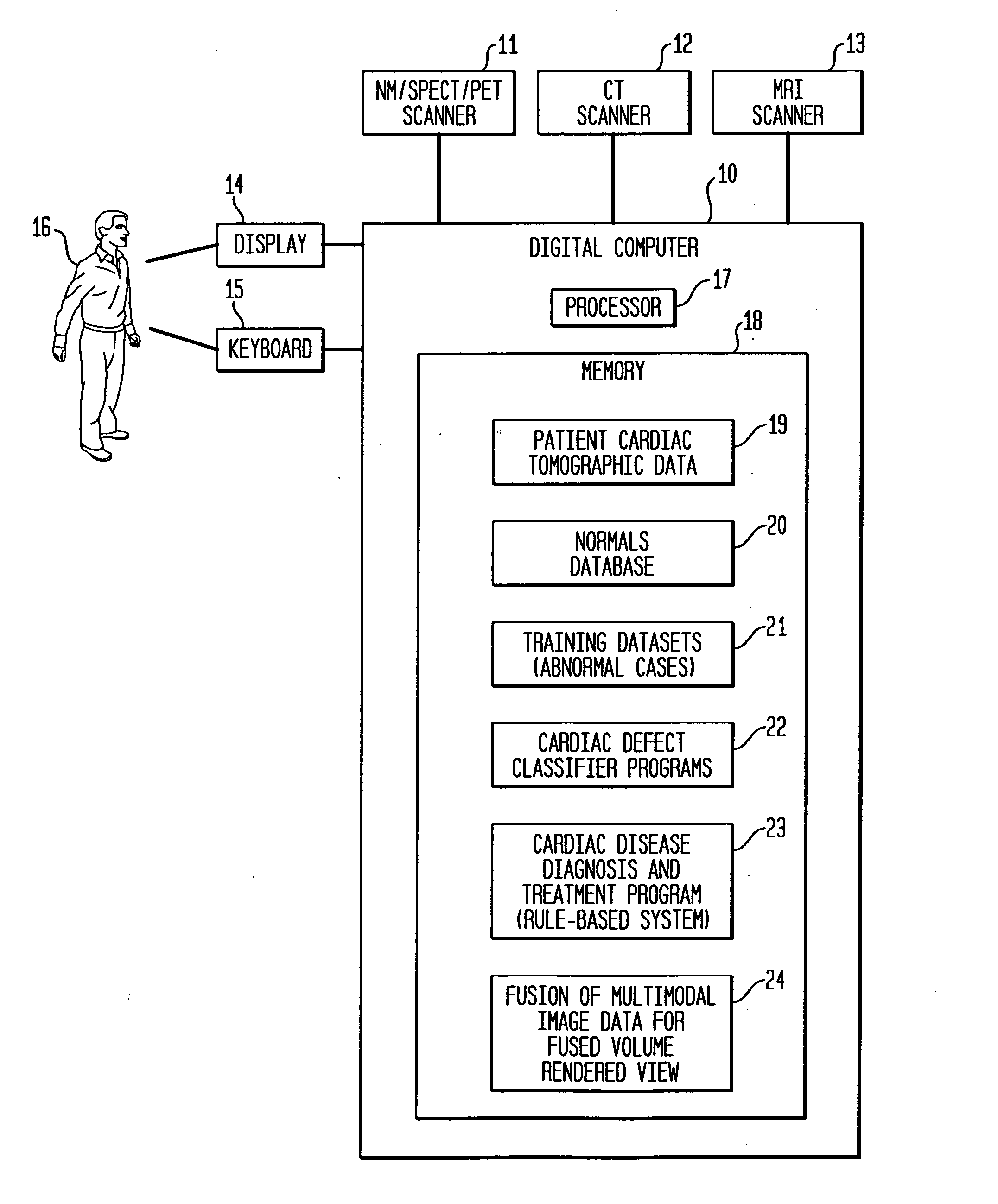
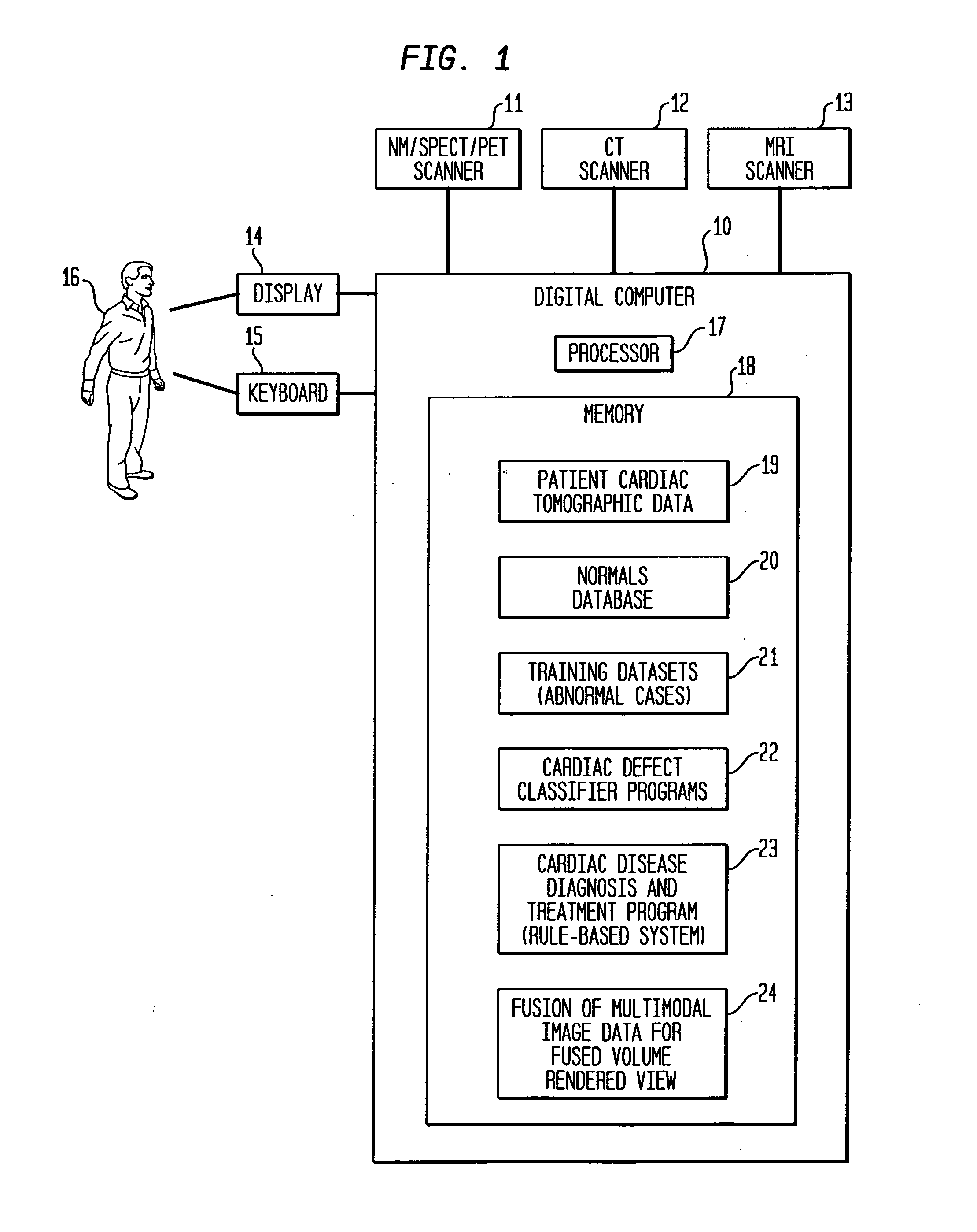
[0015]FIG. 1 shows a system for medical imaging and computer-implemented diagnosis and treatment of cardiac disease. The system includes a digital computer 10 and an NMISPECT / PET scanner 11, a CT scanner 12, and an MRI scanner 13. Additional scanners may be used, such as an ultra-sound (US) scanner. The computer 10 is linked to a display 14 and a keyboard 15 to provide an interface to a human user 16. The computer includes a processor 17 and a memory 18. The memory 18 stores a database 19 of patient cardiac tomographic data from the scanners 11, 12, 13; a normals database 20 of cardiac measurements of healthy patients, and a database 21 of training datasets including abnormal cardiac measurements from patients having cardiac disease. The memory 18 also stores cardiac defect classifier programs 22 for identifying cardiac defects in a patient from the patient cardiac tomographic data 19, and a rule-based cardiac disease diagnosis and treatment program 23 for diagnosing and treating ca...


PUM


Abstract

For diagnosis and treatment of cardiac disease, images of the heart muscle and coronary vessels are captured using different medical imaging modalities; e.g., single photon emission computed tomography (SPECT), positron emission tomography (PET), electron-beam X-ray computed tomography (CT), magnetic resonance imaging (MRI), or ultrasound (US). For visualizing the multi-modal image data, the data is presented using a technique of volume rendering, which allows users to visually analyze both functional and anatomical cardiac data simultaneously. The display is also capable of showing additional information related to the heart muscle, such as coronary vessels. Users can interactively control the viewing angle based on the spatial distribution of the quantified cardiac phenomena or atherosclerotic lesions.

Description

BACKGROUND OF THE INVENTION [0001] 1. Field of the Invention [0002] The present invention relates generally to medical imaging, and more particularly to computer processing of cardiac image data for diagnosis and treatment of cardiac disease. [0003] 2. Description of the Background Art [0004] Medical imaging is one of the most useful diagnostic tools available in modern medicine. Medical imaging allows medical personnel to non-intrusively look into a living body in order to detect and assess many types of injuries, diseases, conditions, etc. Medical imaging allows doctors and technicians to more easily and correctly make a diagnosis, decide on a treatment, prescribe medication, perform surgery or other treatments, etc. There are medical imaging processes of many types and for many different purposes, situations, or uses. They commonly share the ability to create an image of a bodily region of a patient, and can do so non-invasively. Examples of some common medical imaging types are ...

Claims


Application Information

Patent Timeline
IPC IPC(8): G06K9/00
CPCG06F19/321G06T2207/30004G06T3/4061G06F19/345G16H50/20G16H30/20
Inventor DESH, VLADIMIRO'DONNELL, THOMAS
Owner SIEMENS MEDICAL SOLUTIONS USA INC
Why Eureka
Try Eureka