Cationic anti-microbial peptides and methods of use thereof

a technology of cationic antimicrobial peptides and peptides, which is applied in the field of cationic antimicrobial peptides and, can solve the problems of unintended effect of breeding resistant strains of bacteria, severe, even life-threatening infection in the human host, and many symptoms only apparen

Inactive Publication Date: 2006-10-26
WOUNDCHEK LAB US
View PDF10 Cites 5 Cited by
  • Summary
  • Abstract
  • Description
  • Claims
  • Application Information

AI Technical Summary

Problems solved by technology

Infection of wounds is a major source of healthcare expenditure in the United States.
For example, Staphylococci are usually carried as harmless commensals, however given a breach in the epidermis, they can cause severe, even life threatening infection in the human host.
While generally effective, this strategy has the unintended effect of breeding resistant strains of bacteria.
Unfortunately, many symptoms are only evident after the infection is already established.
Furthermore, after a patient is discharged from the hospital they become responsible for monitoring their own healthcare, and the symptoms of infection may not be evident to the unskilled patient.

Method used


Image

  • Cationic anti-microbial peptides and methods of use thereof
  • Cationic anti-microbial peptides and methods of use thereof
  • Cationic anti-microbial peptides and methods of use thereof

Examples


example 1

Detection Assays

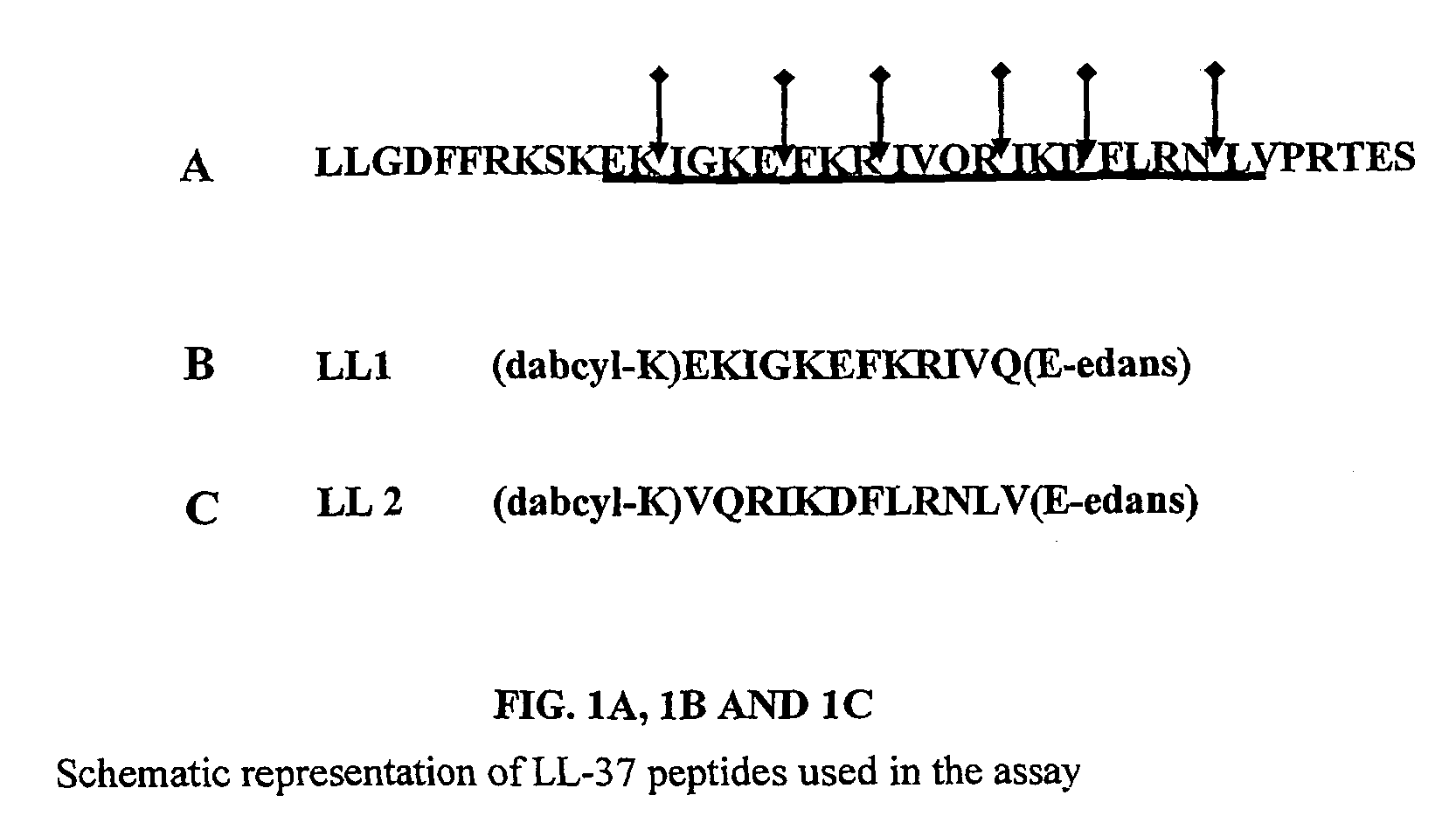
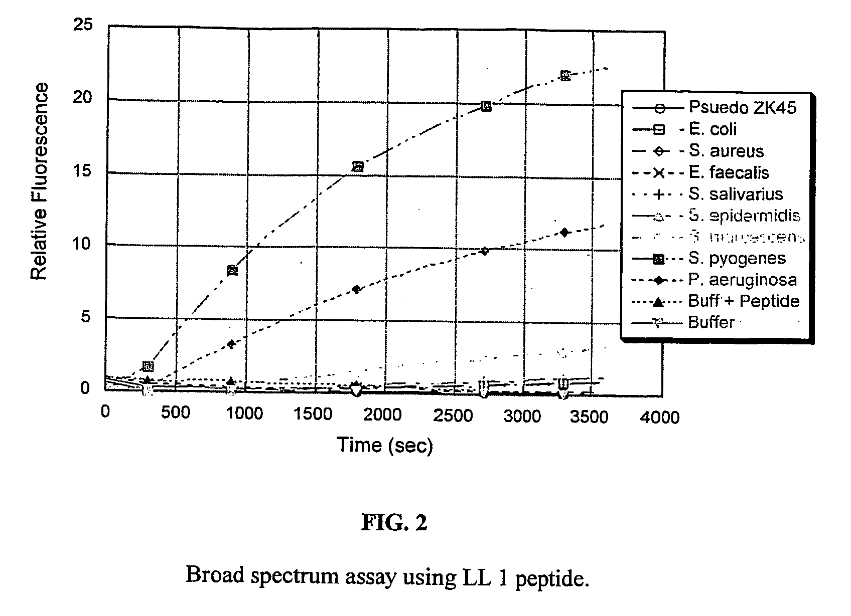
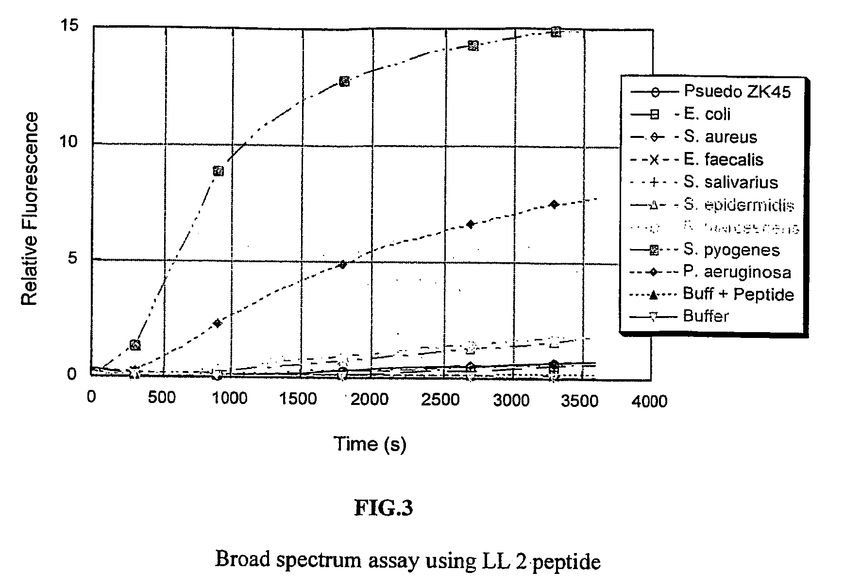
[0063] Two peptide substrates, LL 1 and LL 2, were synthesized so as to encompass all the cleavage sites of the LL-37 as shown in FIGS. 1A, 1B& C. The peptide substrates used here were labeled with the fluorescent probe edans (5-((2-aminoethyl)amino)naphthalene-1-sulphonice acid) and the quencher dye molecule dabcyl((4-(4-(dimethylamino)phenyl)azo)benzoic acid).

[0064] The bacteria were grown in an incubator overnight at 37 C in 5 ml BHI (Brain Heart Infusion) media. Each of the resulting cultures was split into two samples. One was used as is, and the other was spun down and the supernatant was collected. The assays were run in 20 mM tris buffer (pH 7.4) with 150 mM NaCl added. The reaction was carried out with 7 ul of culture or supernatant and 3 ul peptide substrate (5 mg / ml in water) in 100 ul total volume at 37 C. The reaction was followed on a fluorimetric plate reader using an excitation wavelength of 355 nm and an emission wavelength of 485 nm.

[0065] The fi...

example 2

Porcine Wound Fluid Assay

[0071] In order to test for cross reactivity in a wound, the assays were performed under physiologically relevant conditions. The assay for LL1 and LL2 were tested using porcine wound fluid extract in place of reaction buffer.

[0072] Immediately after surgery to create partial thickness wounds for the sensor study a gauze dressing was placed on the wound. After a time the dressing was removed, bagged, and placed in a freezer at −80 C. Absorbed wound fluid was recovered from the dressing by incubation in 10 mls buffer (0.1M Tris-HCl, pH 7.4 with 0.1% Triton X-100). The dressing was incubated on both sides for 2 hours at room temperature and squeezed out. The fluid obtained was aliquoted out and frozen at −20 C.

[0073] The bacteria were grown in an incubator overnight at 37 C in 5 ml BHI (Brain Heart Infusion) media. Each of the resulting cultures was spun down and the supernatant was collected. The assays were run in freshly thawed wound fluid. The reaction ...


PUM

PropertyMeasurementUnit
with wavelengthaaaaaaaaaa
pHaaaaaaaaaa

Abstract

Described herein are methods of detecting a wound infection and for detecting the presence or absence of microorganisms, for example, wound pathogens in a sample, by contacting a sample with a cationic anti-microbial peptide that is degradable by an enzyme produced and / or secreted by a microorganism, and detecting degradation or the absence of degradation of the peptide, as an indicator of the presence or absence of the enzyme in the sample, and thus indicative of the presence or absence of a microorganism in the sample. The present invention also features a biosensor for detecting the presence or absence of a microorganism in a sample.

Description

RELATED APPLICATIONS [0001] This application claims the benefit of U.S. Provisional Application No. 60 / 444,521, filed on Jan. 31, 2003. The entire teachings of the above application are incorporated herein by reference.BACKGROUND OF THE INVENTION [0002] Infection of wounds is a major source of healthcare expenditure in the United States. Approximately 5% of all surgical wounds become infected with microorganisms, and that figure is considerably higher (10-20%) for patients undergoing abdominal surgery. Colonization rates are significantly higher in the hospital setting, both among healthcare workers, and among patients. Moreover, the colonizing organisms in the hospital environment are likely to be resistant to many forms of anti-microbial therapy, due to the strong selective pressure that exists in the nosocomial environment, where antibiotics are frequently used. For example, Staphylococci are usually carried as harmless commensals, however given a breach in the epidermis, they ca...

Claims


Application Information

Patent Timeline
Patent Type & Authority Applications(United States)
IPC IPC(8): C12Q1/04G01N33/543
CPCG01N33/54366C12Q1/04
Inventor SANDERS, MITCHELLCOLPAS, GERARDSEBASTIAN, SHITEELLIS-BUSBY, DIANE
Owner WOUNDCHEK LAB US
Why Eureka
Try Eureka