

Method of screening for drugs that block ligand binding to a lipid binding protein
- Summary
- Abstract
- Description
- Claims
- Application Information
AI Technical Summary
Benefits of technology
Problems solved by technology
Method used
Image


Examples
example 1
[0069] Five identical 96-well plates were prepared with 0.5 μM ADIFAB and 1% dimethyl sulfoxide in 200 μL aqueous buffer (20 mM HEPES, 140 mM NaCl, 5 mM KCl, 1 mM Na2HPO4, pH 7.4) to test assay reproducibility, precision, and quality. Wells were excited at 386 nm and the emission detected at 432 and 505 nm with a fluorescence plate reader connected via fiber optic cables to a standard Spex spectrofluorometer. The emission ratio (505 / 432), which determines the amount of bound ADIFAB, was calculated for each well. A positive control (4 μM sodium oleate (OA)) was added to each well and the ratio was measured again. The average ratio (ρ), standard deviation (σ), coefficient of variation (CV), signal-to-background (S / B) and Z′-factor were calculated for each plate (Table 7). The Z′-factor, as defined below, provides a quantitative assessment of the assay [Zhang, J., et al J. Biomol. Screen. 4, 67-73 (1999)]. Z′=1-3σC++3σC-ρC+-ρC-
[0070] Subscripts C+ and C− refer to positive (with OA) ...
example 2
[0071] A small molecule library was screened for binding to ADIFAB in 96-well plates with a fluorescence plate reader connected via fiber optic cables to a standard Spex spectrofluorometer. Each well contained 200 lL aqueous buffer (20 mM HEPES, 140 mM NacC, 5 mM KCl, 1 mM Na2HPO4, pH 7.4), 0.5 μM ADIFAB, and 5 μM screening compound. Wells were excited at 386 nm and the emission detected at 432 and 505 nm. The emission ratio (505 / 432), which determines the amount of bound ADIFAB, was calculated for each well before and after the addition of screening compound.
[0072] Preliminary hits were detected by comparison of the change in the ratio (ΔR) after addition of the screening compound with that for 4 μM oleate (ΔR=1.8). Table 8 shows the results for an example plate of 80 screening compounds. The largest response from this plate was for well A9 and was significantly greater than the positive control. The affinity of this compound for ADIFAB was confirmed and quantitatively determined ...
example 3
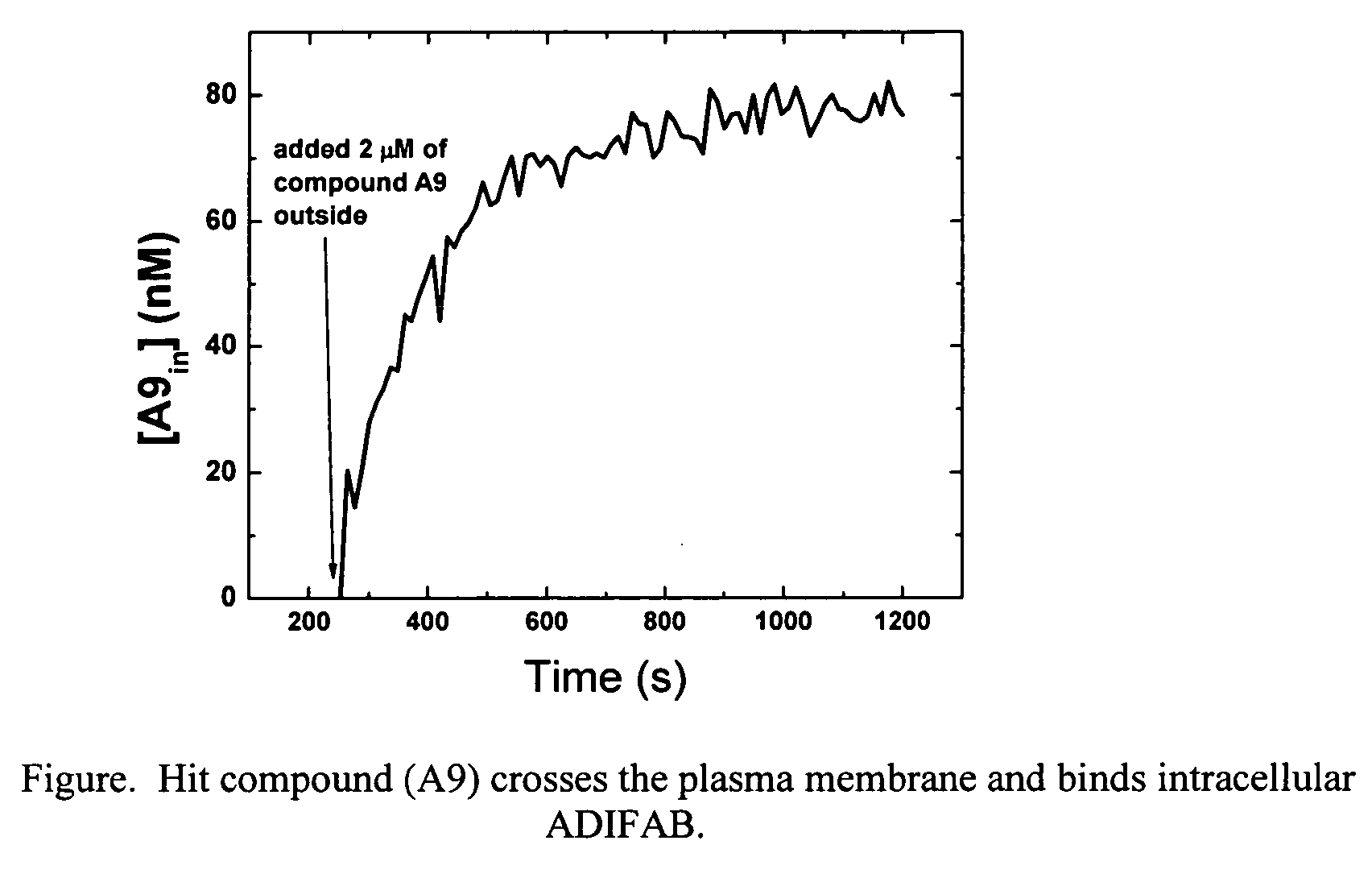
[0073] The plasma membrane permeability of the hit compound (A9) from Example 2 was determined using 3T3-F442A preadipocyte cells loaded with ADIFAB. The syringe-loading technique [Clarke, M. S. F., McNeil, P. L., J. Cell Sci. 102, 533-541 (1992)] was used to introduce ADIFAB into the cytosol of the preadipocytes. A suspension of 105 loaded cells in 1.5 mL aqueous buffer (20 mM HEPES, 140 mM NaCl, 5 mM KCl, 1 mM Na2HPO4, pH 7.4) was prepared in a continuously mixing glass cuvette. The ADIFAB fluorescence emission at 432 and 505 nm was recorded every 12 seconds while exciting at 386 nm using a standard Spex spectrofluorometer. After 252 seconds, 2 tM of compound A9 was added to the extracellular milieu. The ADIFAB emission ratio (505 / 432) increased after the addition of the hit compound indicating that the compound can permeate the plasma membrane and bind to intracellular ADIFAB. The intracellular concentration of compound A9 was calculated from the ADIFAB ratio, and the influx time...
PUM
 Abstract
Description
Claims
Application Information