
Atrial fibrillation detection method and apparatus

a technology of atrial fibrillation and detection method, applied in the field of atrial fibrillation detection method and apparatus, can solve the problems of increased risk of stroke, heart failure, blood clots forming in the atria,

Inactive Publication Date: 2006-12-07
HEWLETT PACKARD DEV CO LP
View PDF13 Cites 38 Cited by
  • Summary
  • Abstract
  • Description
  • Claims
  • Application Information

AI Technical Summary

Benefits of technology

[0014] The present invention allows the electrodes to be placed in any position on the chest wall, that is, it allows changing morphology by allowing positioning of an electrode in a non-standard location.
[0015] In particular, the present invention provides a solution for detecting atrial fibrillation in non-standard lead configurations, in a noisy signal from an ambulatory subject from a sensor rotated through multiple placements. Atrial fibrillation can be detected from a low cost sensor which may be a small form factor sensor with one inch lead separation.
[0016] The nature of the detection technique (method and apparatus), in terms of robustness and ECG sensor device morphology independence, allows it to be used in non-standard electrode lead placements and allows seamless detection as the electrode (sensor) is moved to different positions. This enables the sensor device (electrode) to be placed in a different location each day, which reduces skin irritation in any one particular location, allowing long term wearability. The robustness of the algorithm to non-standard electrode lead placements does not require placement by someone who has received special training in the placement of the electrodes which decreases the cost of the testing and allows the device to be used for preventative care. As the analysis is morphology independent, it does not require a match with a template for a normal trace for a particular position.

Problems solved by technology

AF is not immediately life threatening, but the risk of stroke is increased because the quivering atria beat too rapidly to contract effectively and with time they enlarge, which can lead to blood clots forming within the atria.
If a blood clot leaves the heart and lodges in the brain, a stroke results.
Also, the rapid beating of the ventricles for prolonged periods can result in weakening them which can lead to heart failure.
The ECG signal generated by the ambulatory monitor often contains significant muscle noise as the subject is ambulatory which makes it difficult to detect AF.
Furthermore, due to the limitation in available memory in the ambulatory monitor, only a fixed length recording can be stored, so the individual may be asked to only record when recognizing a rapid heart beat or the device may record over previously recorded data.
The need for a person skilled in placement of the electrodes increases the cost of the ECG and limits the use of the test to those who have already exhibited symptoms.
Long term use, for example, for a time period greater than 24 hours, can result in skin irritation due to exposure to the adhesive.

Method used


Image

  • Atrial fibrillation detection method and apparatus
  • Atrial fibrillation detection method and apparatus
  • Atrial fibrillation detection method and apparatus

Examples


Embodiment Construction

[0032] A description of preferred embodiments of the invention follows.

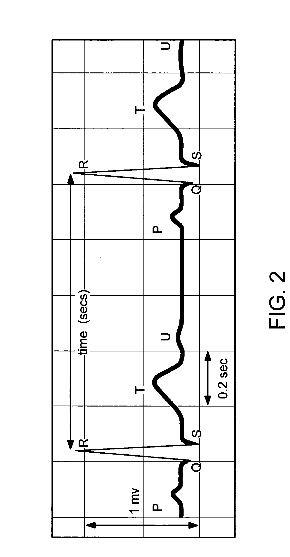
[0033] Most ECG recordings contain two or more simultaneously recorded ECG signals, called “leads.” The heart generates an electrical field that varies spatially as well as temporally. Thus, the standard practice is to record two or more signals (leads) derived using sensing electrodes placed at certain specific locations. The wires that connect the electrodes to the recording equipment are also sometimes referred to as “leads”.

[0034] As is well-known in the art, there is a standard placement for ECG leads that requires an individual with special training to perform the placement. Typically, a nurse performs the placement, a doctor performs the testing, an ECG technician runs the analysis software and a cardiologist performs the over-read. Non-ambulatory electrocardiograph devices include precordial leads and limb leads. Precordial leads are placed on the chest wall at pre-defined positions on the chest wall re...


PUM


Abstract

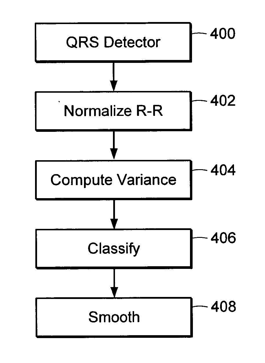
A method for automatically detecting atrial fibrillation in a non-standard ECG signal having changing morphology and containing significant muscle noise generated by an ambulatory subject is provided. A morphology independent QRS detector is used to compute R-R intervals in the ECG signal. The variance of the R-R intervals over a sliding window is normalized and compared with a threshold to determine if atrial fibrillation is present within the window.

Description

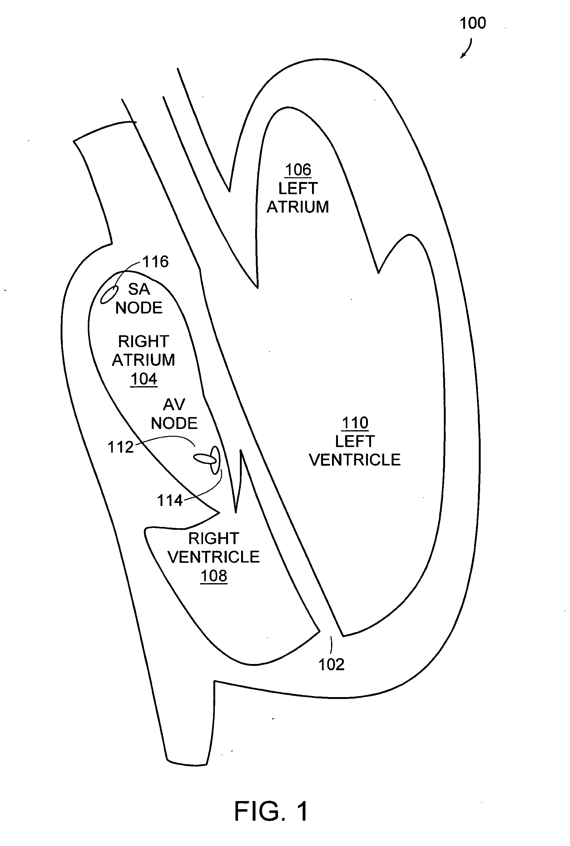
BACKGROUND OF THE INVENTION [0001] As shown in FIG. 1, the heart 100 is a pump, has four chambers and is divided into a right side and a left side by a muscular wall called the septum 102. The two chambers at the top are called the right atrium 104 and the left atrium 106 and the two chambers at the bottom are called the right ventricle 108 and the left ventricle 110. The atria and ventricles work together, contracting and relaxing to pump blood out of the heart. [0002] Oxygen-poor blood enters the top of the heart through the inferior and superior vena cava veins and flows into the right atrium 104 and passes through the tricuspid valve to the right ventricle 108. After the right ventricle 104 fills, it contracts and blood flows through the pulmonary valve to the lungs. Oxygen-rich blood from the lungs enters the left atrium 106 through the pulmonary vein and through the left atrium 106 to the left ventricle 110. The left ventricle pumps the blood into the aorta. [0003] The heart a...

Claims


Application Information

Patent Timeline
Patent Type & Authority Applications(United States)
IPC IPC(8): A61B5/04
CPCA61B5/046A61B5/361
Inventor HEALEY, JENNIFERLOGAN, BETH T.
Owner HEWLETT PACKARD DEV CO LP
Learn More