Multi-spectral imaging endoscope system

a multi-spectral imaging and endoscope technology, applied in the field of surgical devices, can solve the problems of inconvenient use of endoscopes, inability to capture and process internal images in real time, and inability to use conventional imaging techniques such as x-ray and mri in conjunction with endoscopes,

Inactive Publication Date: 2007-02-15
MICRO INVASIVE TECH
View PDF11 Cites 18 Cited by
  • Summary
  • Abstract
  • Description
  • Claims
  • Application Information

AI Technical Summary

Benefits of technology

[0013] A further object of the present invention is to provide barrier elements that operatively isolate portions of the endoscope from contact with the patient and / or the user.

Problems solved by technology

However, there is no conventional means for real-time capture and processing of internal images during an endoscopic surgical procedure.
In addition, conventional imaging techniques such as X-ray and MRI are not suitable for use in conjunction with endoscopes.
However, typical endoscopes do not transmit deep into the ultra violet region of the light spectrum because of, among other things, the use of fused silica as the transmitting medium.
Current endoscopes cannot readily combine visual imaging and therapeutic intervention because their light source must be continuous; their fiber optic bandwith is limited; and their optics are inefficient, responding only to light between 400-700 nm.

Method used


Image

  • Multi-spectral imaging endoscope system
  • Multi-spectral imaging endoscope system
  • Multi-spectral imaging endoscope system

Examples


Embodiment Construction

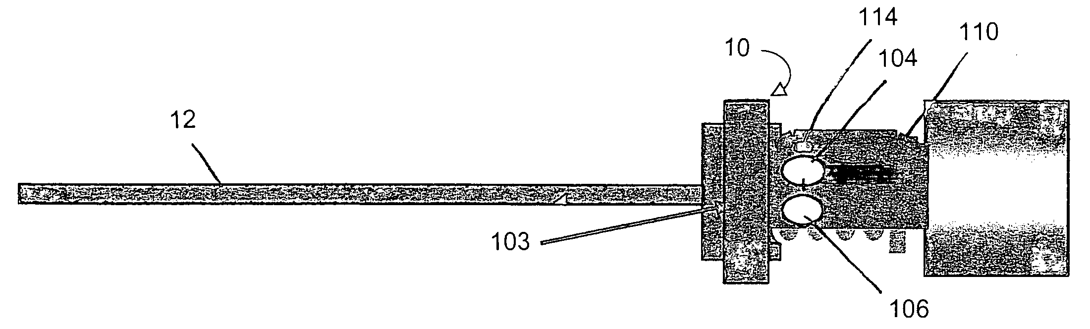
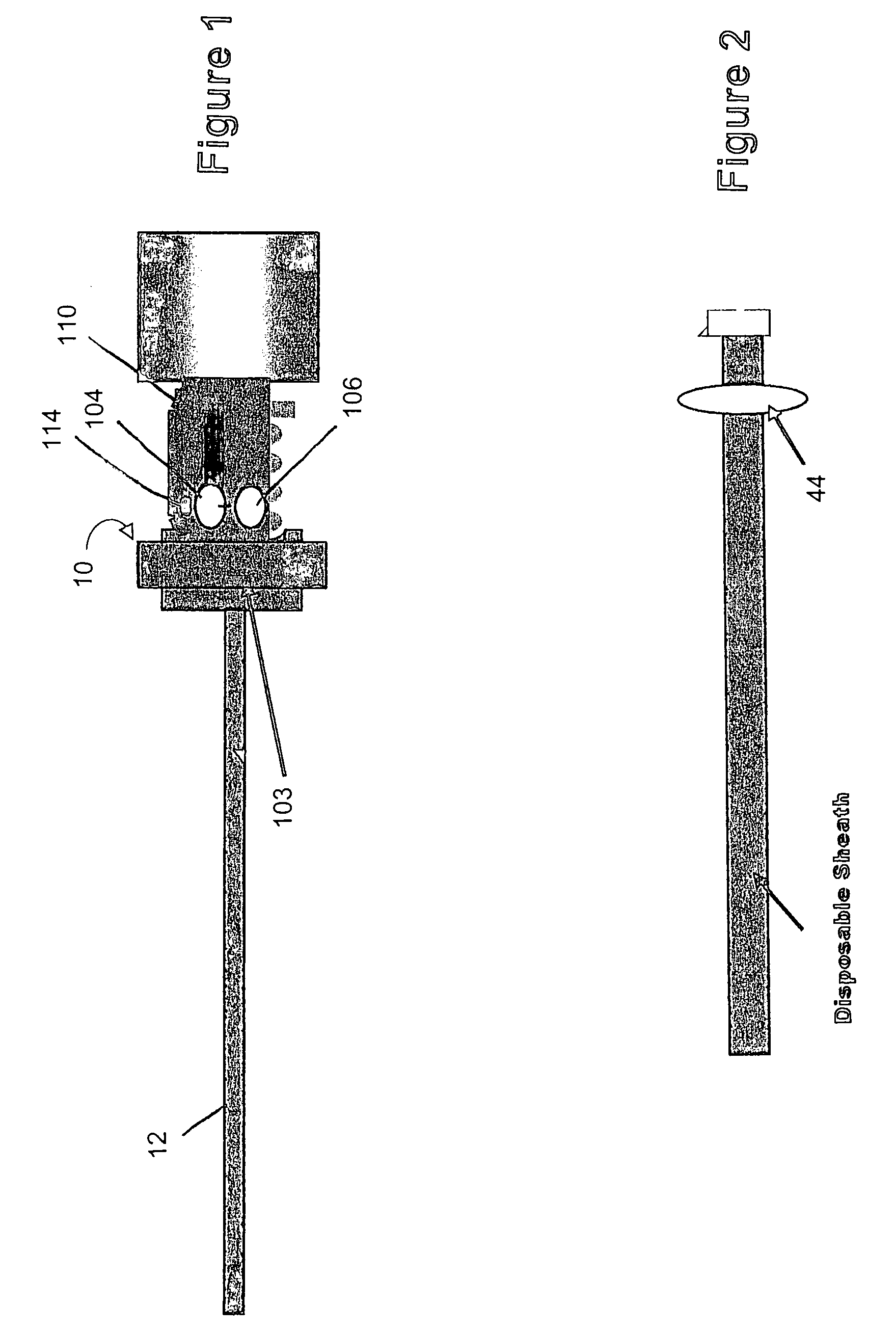
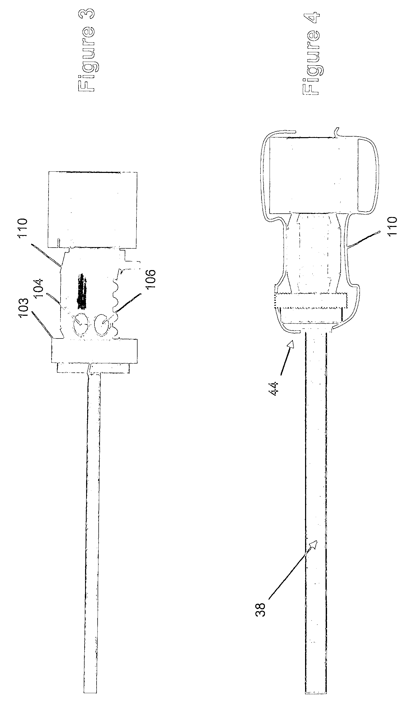
[0026] With reference to the drawings wherein like numerals represent like parts throughout the several figures, a multi-spectral endoscope in accordance with the invention is designated by the numeral 10. The multi-spectral endoscope 10, as shown in FIG. 4, can be used for medical bioimaging within, for example, a patient's abdominal cavity to enhance the visualization of areas of interest. For example, a site of interest located within a patient's abdominal cavity is illuminated with UV light. The site of interest can also be associated with structure, tissue, or fluid which when illuminated with UV light can be distinguished from the surrounding field of view. In addition, UV dye can be used in conjunction with the UV light illumination. The UV dye can, for example, be locally or systematically injected into the patient in order to image structures of interest. The multi-spectral feature refers to illumination in at least the UV and the visual (VIS) ranges.

[0027] In one embodime...


PUM


Abstract

Light is introduced into the body through an endoscope illumination system, which is capable of passing both UV and visible radiation through an illumination pathway. An image can then be viewed in real time, by eye or with an electronic imaging camera and displayed on a video monitor used by the surgeon. Dyes which are activated by the UV radiation generate images that can be viewed by the endoscope in the visible spectrum and recorded by eye, electronic camera or other recording devices that can process visual images.

Description

FIELD OF THE INVENTION [0001] The present invention generally relates to surgical devices. In particular, the invention relates to an endoscope having therapeutic interventional capability combined with imaging elements. BACKGROUND OF THE INVENTION [0002] The ability to view interior portions of a patient's body during a surgical medical procedure is invaluable for efficacious surgical intervention. Conventionally, devices for viewing the interior of a patient's body during a surgical procedure utilize light guides. These conventional light guides allow areas within a patient's body cavities to be both illuminated and visualized through an eyepiece. These conventional systems utilize continuous (CW) light sources that are coupled to an illumination conduit by a light guide and an optical connector located at or near the top of the illumination device. The designers of light sources for use in conventional systems are typically concerned with only light in the visible wavelengths. [0...

Claims


Application Information

Patent Timeline
Patent Type & Authority Applications(United States)
IPC IPC(8): A61B6/00
CPCA61B1/00165A61B1/07A61B1/0638
Inventor BALA, JOHN L.
Owner MICRO INVASIVE TECH
Why Eureka
Try Eureka