Method for navigating a virtual camera along a biological object with a lumen

a technology of biological objects and virtual cameras, applied in the field of virtual cameras along biological objects with lumens, can solve the problems of difficult to give a high-quality real-time viewing experience, and achieve the effect of faster endoscopic examination

Active Publication Date: 2007-03-08
TOSHIBA MEDICAL VISUALIZATION SYST EURO
View PDF11 Cites 76 Cited by
  • Summary
  • Abstract
  • Description
  • Claims
  • Application Information

AI Technical Summary

Benefits of technology

[0009] Navigation performed in accordance with this method will generally avoid the wall of the object, and hence be useful for a range of applications. For example, virtual endoscopy requires navigation for a virtual camera that stays close to the centerline of the object, both to give a good field of view and to avoid giving a user the impression that the camera is about to collide with the object wall. The wall avoidance is achieved by using the direction of a group of rays with the longest average ray length out of a number of groups of rays as the segment direction for the next navigation segment. Selection of the longest individual ray from the segment start point gives a navigation direction generally along the length of the object, but which tends to pass very close to the wall at bends. Replacing this with an average length from the selected group of rays tends to bias the navigation direction away from the wall and towards the centerline, to give an improved navigation. A group of rays having an axial direction that passes close to the object wall at a bend will have a reduced average length (because some rays will meet the wall before the bend and hence be short) compared to a group with an axial direction closer to the centerline. Thus the latter group will be selected to give the segment direction, keeping the navigation near to the centreline.
[0025] The camera may travel at a velocity that can be adjusted by a user. This further contributes to user interaction, allowing the user to travel slowly through regions of the object of most interest to obtain a better view, and to travel quickly through parts of the object of little or no interest, thus allowing an endoscopic examination to be performed more rapidly.

Problems solved by technology

To navigate the lumen, a path for the camera to follow is often determined wholly or partially in advance, since even an experienced clinician may have difficulty in manually controlling the camera movement.
This approach offers some user interaction as the camera progresses along the lumen but it is more difficult to give a high-quality real-time viewing experience owing to the time needed to produce the images.

Method used


Image

  • Method for navigating a virtual camera along a biological object with a lumen
  • Method for navigating a virtual camera along a biological object with a lumen
  • Method for navigating a virtual camera along a biological object with a lumen

Examples


Embodiment Construction

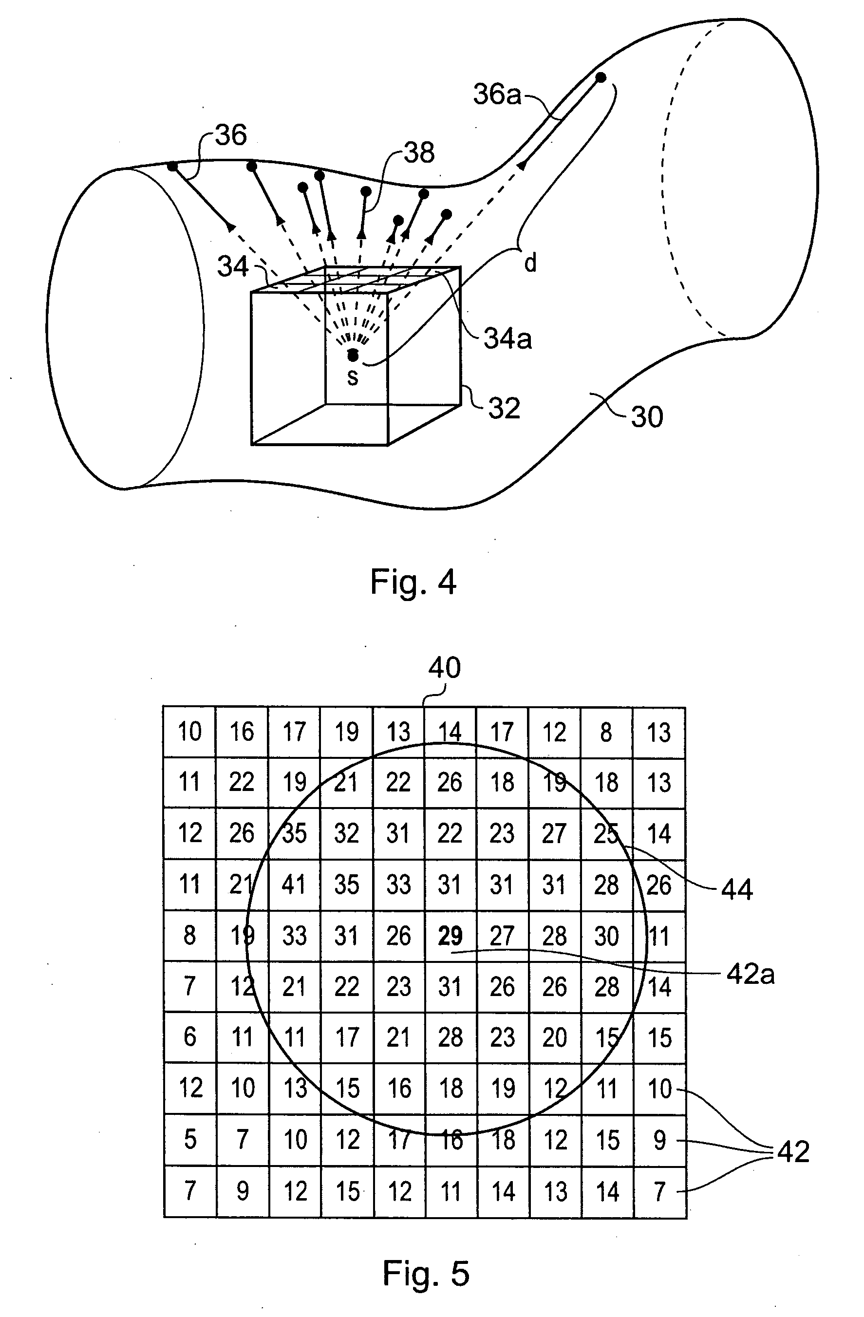
[0045] Medical imaging techniques such as computer-assisted tomography (CT), magnetic resonance imaging (MRI), ultrasound and positron-emission tomography (PET) produce large sets of data representing all or part of a patient's body. The data are typically collected in slices or layers through the patient's body, which are then assembled into a three-dimensional volume data set. The data set comprises a plurality of volume elements or voxels, arranged in a regular three-dimensional array. Each voxel has a value associated with a physical property (expressed in Hounsfield units, for example) that indicates the characteristics of the tissue in the corresponding part of the patient. Thus, voxels representing bone will have different values from voxels representing organs or muscles. Volume rendering, surface rendering or other image processing methods can be used to generate two-, three- or four-dimensional images of the patient from any viewpoint inside the data set.

[0046] One partic...


PUM


Abstract

A method of navigating along a biological object with a lumen represented by a three-dimensional volume data set comprises generating a plurality of navigation segments connectable in a sequence, each segment having a start point within the lumen, a direction and a length. The navigation may be used for a camera in a virtual endoscopic examination, for example. The direction of each segment is determined by casting groups of rays outwards from the start point of the segment to the object wall, and calculating an average ray length for each group. The group having the largest average ray length is selected, and the axial direction of this group is used as the direction for the segment. The average ray lengths of the groups may be weighted using the direction of the previous segments to bias the navigation generally forward, or may be weighted using a view direction of the camera to allow a user to turn the camera into a chosen branch in the object.

Description

BACKGROUND OF THE INVENTION [0001] The present invention relates to a method of navigating along a hollow tubular object such as a biological object with a lumen, the object included in a three-dimensional volume data set representing a biological structure. [0002] Patient imaging methods, such as computer-assisted tomography (CT), magnetic resonance imaging (MRI), ultrasound and positron-emission tomography (PET), generate large three-dimensional volume data sets representing all or part of a patient's body. These volume data sets are highly detailed and allow complex studies of the body to be made. Techniques for this include virtual endoscopy, in which a virtual camera is made to travel along a biological object with a lumen such as a colon, blood vessel or bronchial tree, with the image of the lumen that is seen by the camera presented to a user for diagnostic and other purposes. To navigate the lumen, a path for the camera to follow is often determined wholly or partially in ad...

Claims


Application Information

Patent Timeline
Patent Type & Authority Applications(United States)
IPC IPC(8): G09G5/00
CPCG06T2210/41G06T19/003
Inventor GRAHAM, ALANSMOUT, ANDREWDAVEY, ROBERT A.LESSIEUX, LAURENT
Owner TOSHIBA MEDICAL VISUALIZATION SYST EURO
Why Eureka
Try Eureka