
Method for determining the dispersion behavior of a contrast agent bolus

a contrast agent and dispersion behavior technology, applied in the field of dispersion behavior determination of contrast agent bolus, can solve the problems of difficult evaluation of recorded three-dimensional images, unsatisfactory results can be produced, etc., and achieve the effect of being ready to evalua

Inactive Publication Date: 2007-03-08
SIEMENS AG
View PDF4 Cites 45 Cited by
  • Summary
  • Abstract
  • Description
  • Claims
  • Application Information

AI Technical Summary

Benefits of technology

[0020] Performing subtraction angiography, which is advantageously performed as digital subtraction angiography, it also being possible, of course, to perform analog subtraction angiography, is recommended for the situation in which static regions or, as the case may be, objects in the or of the object under investigation are to be the subject of the images. Cranial images should be mentioned here by way of example. The recorded images can be stored in digital form and subsequently the image objects which are free of contrast agent can be subtracted from the image objects that show contrast agent, thereby producing, as a result of this subtraction, a representation that can be readily evaluated.
[0021] In the case of non-static objects as part of the recorded images, in the area of the heart for example, native images can be taken as preparatory images. Said native images can be evaluated directly, without need for processing by subtraction or the like. Although the native images may be more difficult to evaluate, they avoid errors due to movements of the recorded objects or, as the case may be, areas of the body.
[0024] According to the invention the differences in the determined times t1, t2 and t3 are stored in a control device for the rotational angiography. There they are available directly for setting for the three-dimensional imaging to be performed subsequently. In addition it is of course possible to store the differences of the determined times or the times themselves directly in a central memory device for example for a plurality of examination equipment of a clinic or the like or to use an external storage medium such as a CD-Rom or the like. Storing the data in the control device for a rotational angiography procedure has the advantage that the data can be stored in such a way that a corresponding program means for performing the examination can locate the time data without further processing and initiate the corresponding setting of the C-arm system provided for recording the image.
[0028] In addition, further preparatory images for determining the time curve for the uptake of the contrast agent bolus in the vascular system can be generated and, as a function thereof, at least one further time for recording a rotational angiography image of at least one further phase, in particular a phase of the liver, can be determined and incorporated into the determination of the differences. Thus, a three-phase recording of the liver is recommended for particular medical questions in which an early arterial phase, a portal-venous phase and a late arterial phase are distinguished. Multiphase examinations of this type enable reliable location of lesions with the most varied blood supply types. In such a case a further time, for example a time t4 for an arterial late phase, must be determined in the course of the preparatory images. It is, of course, conceivable to determine further times in addition and include them in the forming of the differences. By determining the differences and programming these delays into a control device for the rotational angiography it is possible for the subsequent images to be taken at their respective optimal times or, as the case may be, in periods specified thereby.
[0031] As already explained, in order to perform the subsequent rotational angiography the determined times t1, t2 and t3 and / or the stored differences can be input via a user interface of a control device. The user interface enables the operator to interact with a program means which controls the data acquisition in order to make changes if necessary, such as for example the input of the individual times determined here for recording the data of the individual phases, so that in contrast to a possibly present standard protocol an optimal image quality of the recorded images can be achieved.

Problems solved by technology

If said times are estimated incorrectly, unsatisfactory results can be produced in the course of the rotational angiography procedure, with images on which the separation between the arterial and the venous system in particular is not clear.
As a consequence the evaluation of the recorded three-dimensional images is rendered more difficult.

Method used


Image

  • Method for determining the dispersion behavior of a contrast agent bolus
  • Method for determining the dispersion behavior of a contrast agent bolus
  • Method for determining the dispersion behavior of a contrast agent bolus

Examples


Embodiment Construction

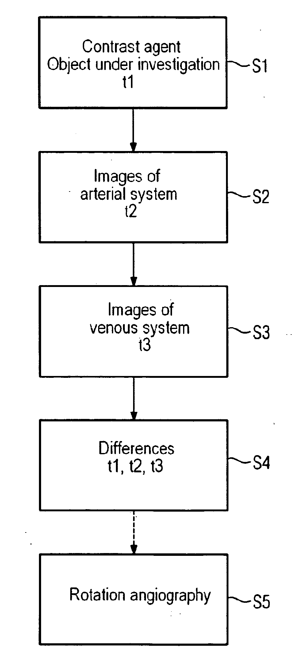
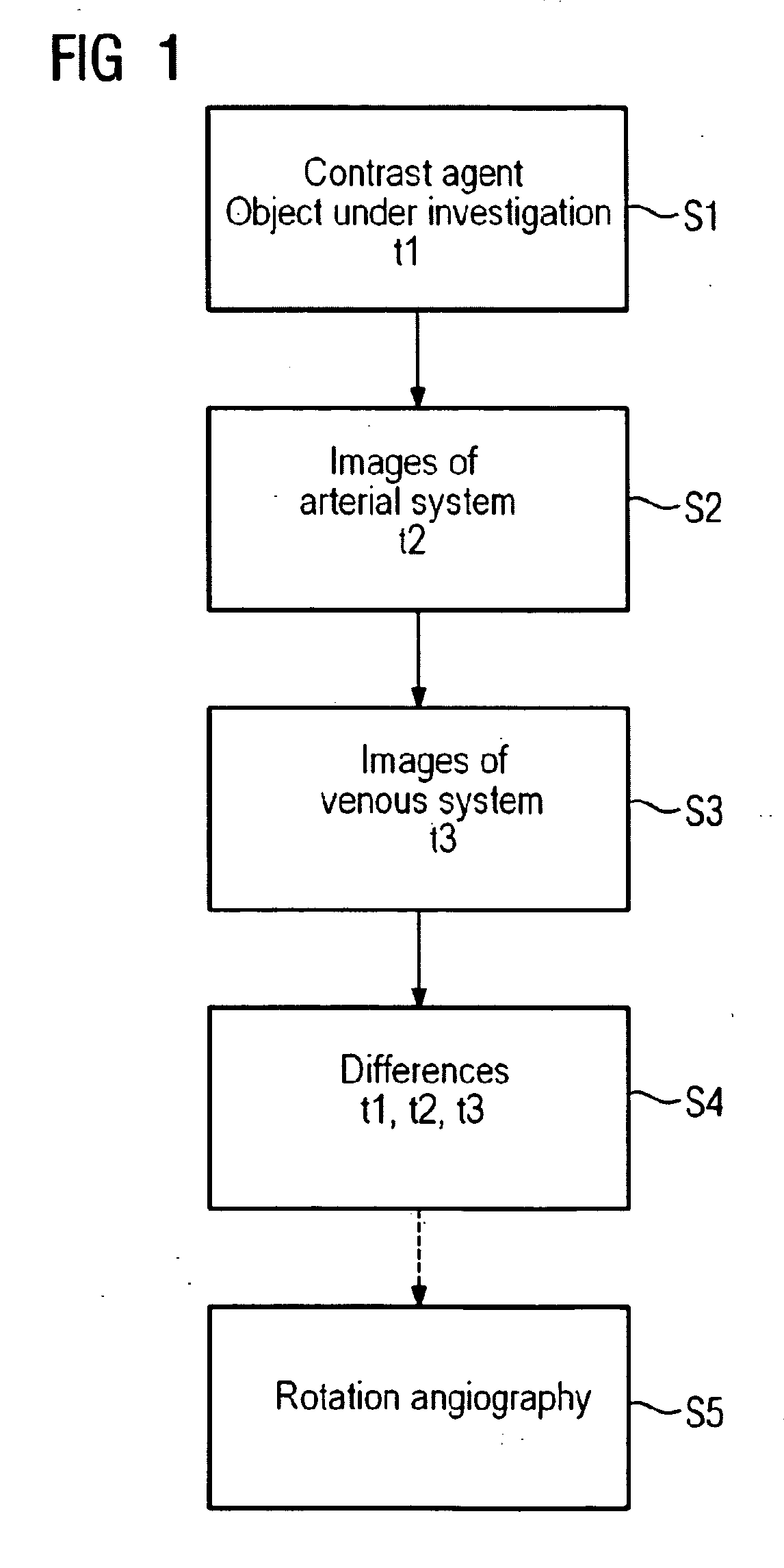
[0039]FIG. 1 shows a flow diagram of a method according to the invention. Here, in order to determine the dispersion behavior of a contrast agent bolus in the blood vessel system, contrast agent is first introduced into same. This takes place at time t1 with the aid of a suitable injector.

[0040] Next, in step S2, preparatory images are produced as test images which show at least a part of the vascular system of the object under investigation in order to reveal the time curve of the uptake of the contrast agent bolus in the arterial vascular system. From the influence of the contrast agent on the recorded images through discoloration and the like, a time t2 for recording a rotational angiography image of an arterial phase is determined, said time being specified by the uptake in the arterial vascular system. If appropriate, test images of different arterial phases can be produced, for example of an early arterial and a late arterial phase.

[0041] Preparatory images are also produced...


PUM


Abstract

A method for determining the dispersion behavior of a contrast agent in the blood vessel system of an object for a rotational angiography procedure, comprising: introducing the contrast agent into the blood vessel system and determining the time t1 of said introduction, producing preparatory images of at least a part of the vascular system in order to determine the time curve for the uptake of the contrast agent in the arterial vascular system and, determining a time t2 for recording a rotational angiography image of an arterial phase, producing preparatory images of at least a part of the vascular system in order to determine the time curve for the uptake of the contrast agent in the venous vascular system and, determining a time t3 for recording a rotational angiography image of a venous phase, and determining the differences in the times t1, t2 and t3 for the rotational angiography.

Description

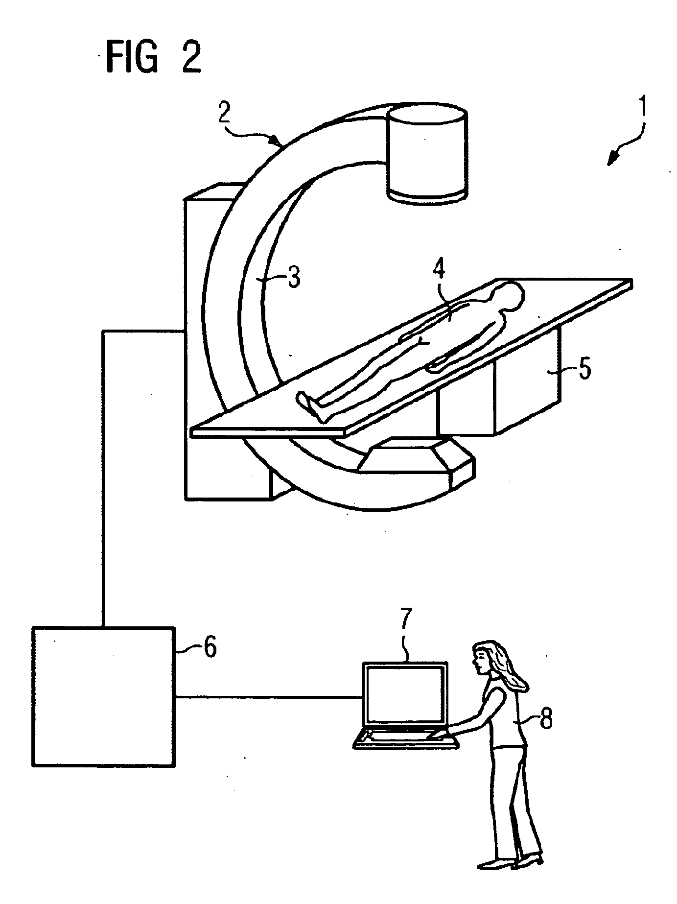
CROSS REFERENCE TO RELATED APPLICATIONS [0001] This application claims priority of German application No. 10 2005 042 328.0 filed Sep. 6, 2005, which is incorporated by reference herein in its entirety. FIELD OF THE INVENTION [0002] The invention relates to a method for determining the dispersion behavior of a contrast agent bolus in the blood vessel of an object under investigation for a rotational angiography procedure that is to be performed. BACKGROUND OF THE INVENTION [0003] Modern C-arm systems enable a three-dimensional representation of vascular trees to be obtained. Toward that end, the C-arm is rotated around the patient in order to record two-dimensional projections from which the anatomical structure of interest is subsequently reconstructed three-dimensionally. For this purpose mathematical algorithms are used by means of which it is possible, as part of what is referred to as cone-beam computer tomography, for example, to determine the three-dimensional reconstruction....

Claims


Application Information

Patent Timeline
IPC IPC(8): A61B6/00
CPCA61B6/504A61B6/481
Inventor KLINGENBECK-REGN, KLAUS
Owner SIEMENS AG
Learn More