Trocar-cannula complex, cannula and method for delivering biologically active agents during minimally invasive surgery

a technology of biological active agents and cannulas, applied in trocar, surgery, medical science, etc., can solve the problems of increased operative time, contamination of the port site during surgery, and increased exposure of the surgical staff to needle sticks, so as to reduce or eliminate the

Inactive Publication Date: 2007-03-29
RXTROCAR +1
View PDF71 Cites 31 Cited by
  • Summary
  • Abstract
  • Description
  • Claims
  • Application Information

AI Technical Summary

Benefits of technology

[0017] As alternative embodiments, the outer layer may be comprised of a biodegradable component that contains an active agent which is time released. The outer layer may be configured similar to a condom and rolled onto the cannula and which includes the necessary aperture(s) and / or channels for delivery of the active agent to the patient from a medical pump or syringe. The outer layer may be a rigid layer which is coupled to the inner cannula member in a rigid fashion or, for example, in a movable fashion such as a rotatable fashion to allow opening, closing, or size adjustment of the delivery passage(s). As one additional alternative, the outer layer may be formed at least partially of a porous material which provides the necessary apertures. Such porous materials may, for example, take the form of sintered metals, filter media, paper, mesh cloth or a porous plastic. The porous material may also be formed of or contain a biodegradable matrix that entraps at least one biologically active agent. As the matrix degrades, the agent is released in a controlled manner resulting in a sustained effect of the agent over a period of time.
[0018] Another embodiment of the invention provides an expandable sleeve that may itself comprise a cannula through which a trocar or trocar assembly is inserted or which may take the place of the perforated outer layer of the grooved cannula discussed above. Other expandable sleeve embodiments may also be configured in accordance with this aspect of the invention as well. Such an expandable sleeve can, for example, allow trocars having different diameters to be inserted through the sleeve. Therefore, the same expandable fluid delivery sleeve may be used in connection with different sized trocars or trocar assemblies thereby reducing or eliminating the need for different sized fluid delivery cannulas or sleeves. The sleeve may also be associated with a biodegradable matrix that contains one or more biologically active agents that are time released as the matrix degrades.

Problems solved by technology

The technique, however, also resulted in increased operative time and increased exposure of the surgical staff to needle sticks.
In addition, the potential for contaminants to spread to the port site during the surgery has been well documented.
Irrigation performed only at the end of the surgical procedure unfortunately cannot reduce patient exposure to contaminants during the procedure.
These stages result in the exacerbation of the pain cascade.
The patient, who is under general anesthesia, is not able to withdraw from the painful stimulus.
There are, however, several problems with this technique.
Second, the injection is in an extrapolated direction of where the obliquely inserted trocar's path lies and, therefore, the anesthetic may not be accurately delivered to the trocar damaged area.
Delivery of the local anesthetic by a syringe and needle also adds to the number of surgical steps to be performed and increases the chances of needle sticks.
As a result of the necessity to deliver the medication via the needle and syringe, and the fact that the medication is short acting in its nature, the patient frequently experiences “break through pain” at the port site.

Method used


Image

  • Trocar-cannula complex, cannula and method for delivering biologically active agents during minimally invasive surgery
  • Trocar-cannula complex, cannula and method for delivering biologically active agents during minimally invasive surgery
  • Trocar-cannula complex, cannula and method for delivering biologically active agents during minimally invasive surgery

Examples


Embodiment Construction

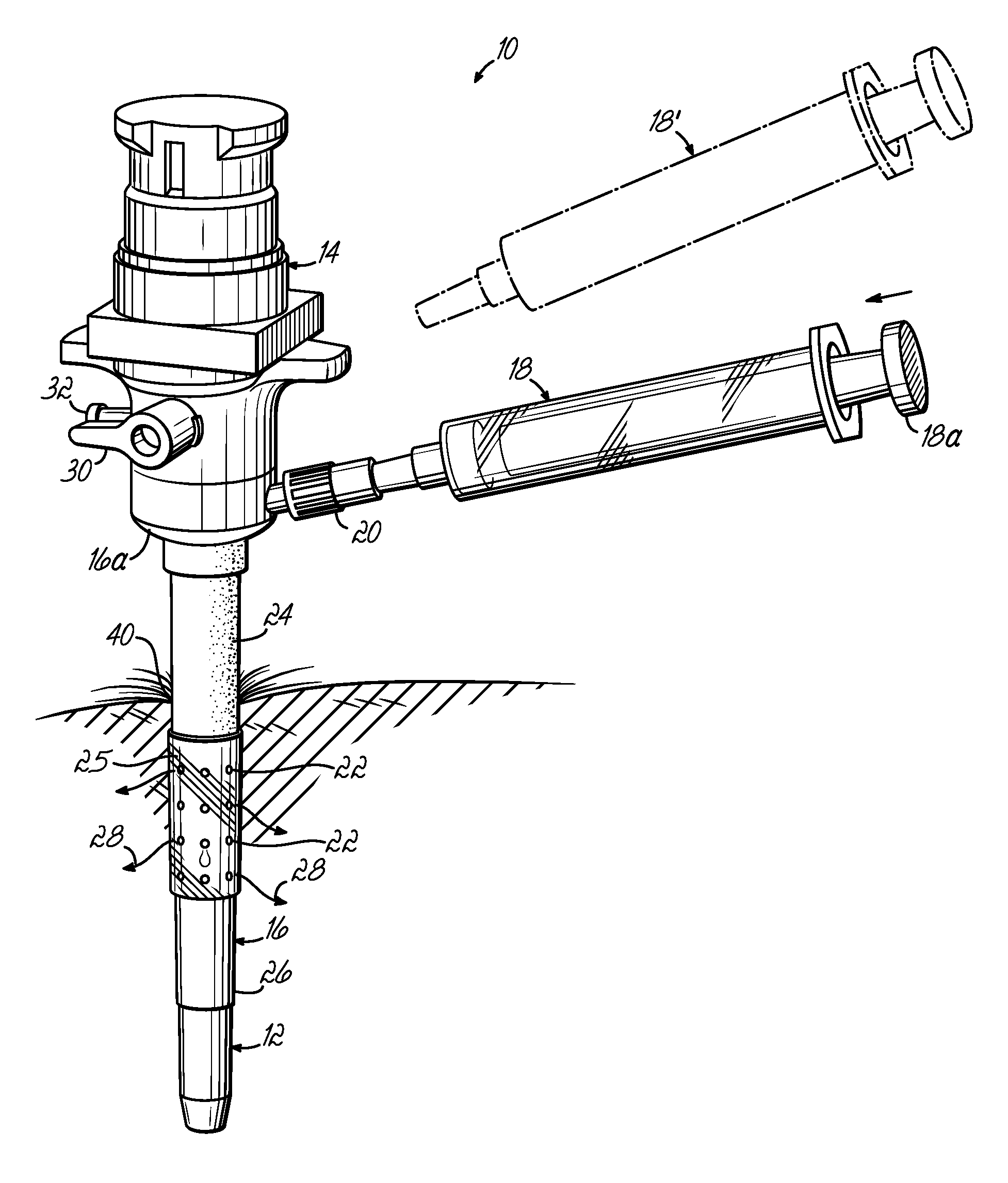
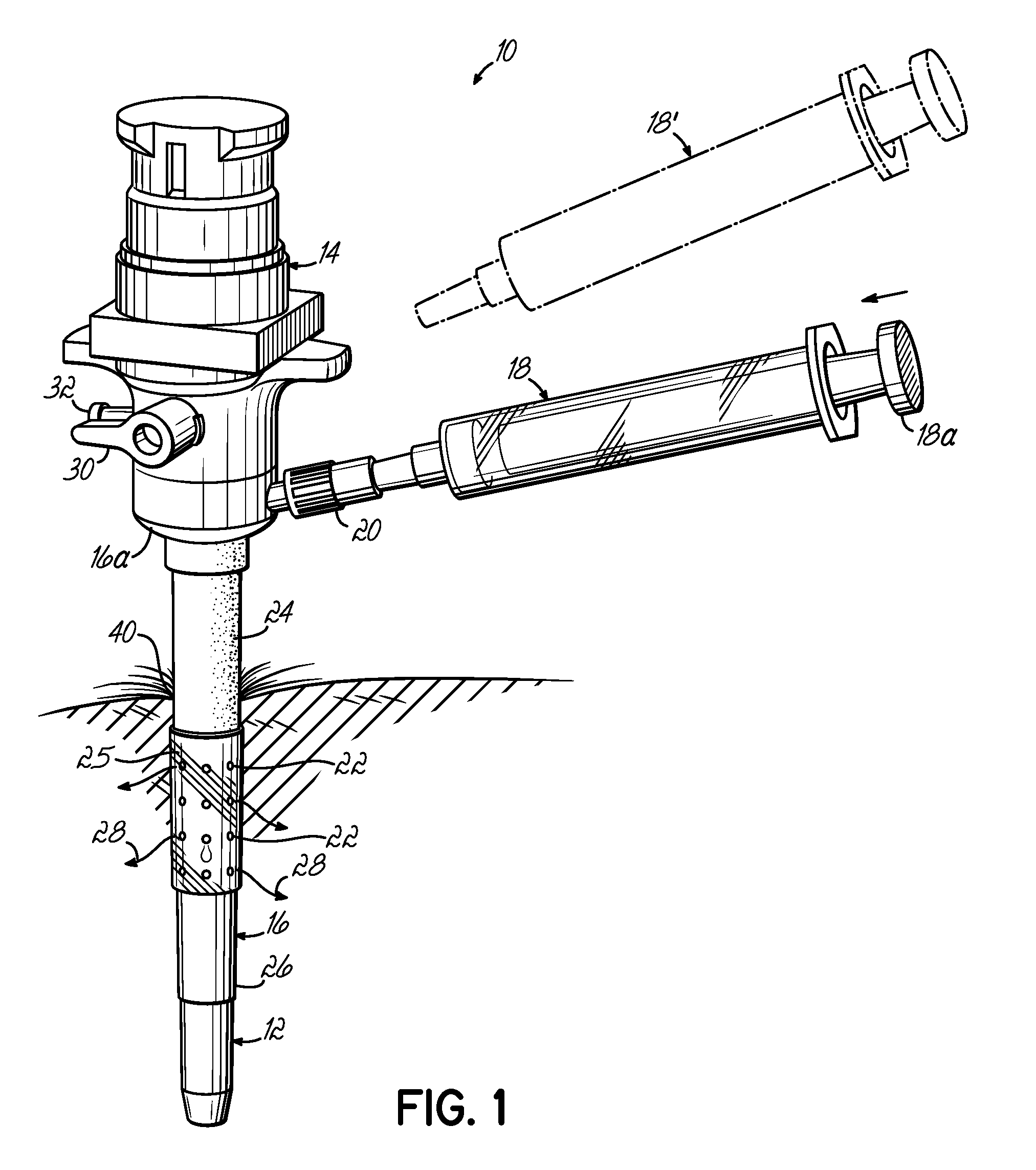
[0031]FIG. 1 illustrates a trocar-fluid delivery cannula complex 10 constructed in accordance with one preferred embodiment of the invention. Complex 10 includes a trocar assembly 12 which may include a conventional hub assembly 14. Representative trocar assemblies are shown and described in previous patents, such as my previous U.S. Pat. Nos. 6,063,060; 6,039,725; 5,865,817; and 5,865,809, and PCT Application No. PCT / US02 / 29356, the disclosures of which are hereby fully incorporated by reference herein. The present invention may implemented into cannulas associated with various other minimally invasive procedures including, but not limited to, laparoscopic procedures.

[0032] In accordance with the invention, a cannula 16 is positioned on the outside of trocar assembly 12 and includes a base portion 16a. A syringe 18 couples to base portion 16a of cannula 16 through a fluid coupling, such as a standard luer connector assembly 20. A second syringe 18′ may be provided to, for example,...


PUM


Abstract

A trocar-cannula complex for use in minimally invasive surgical procedures performed through a port site of a patient and in the delivery of biologically active agents to the patient includes a trocar and a cannula. The cannula includes a tubular structure with a central lumen receiving the trocar and an outer surface adapted to interface with tissue at the port site. A first delivery mechanism is associated with the cannula for delivering a first biologically active agent to a patient and a second delivery mechanism is associated with the cannula for delivery of a second biologically active agent to the patient. Various other manners of delivering the agents are also disclosed.

Description

[0001] This application is a continuation of PCT Serial No. PCT / US2005 / 007678 filed Mar. 8, 2005 which claims the priority of U.S. Provisional Application No. 60 / 552,048, filed Mar. 10, 2004, the disclosures of which are fully incorporated by reference herein.FIELD OF THE INVENTION [0002] This invention generally relates to cannulas and, more specifically, to cannulas used during minimally invasive surgery for allowing the introduction of instruments, such as laparoscopic tools, during surgical procedures and for the delivery of biologically active agents to the port site of the patient. BACKGROUND OF THE INVENTION [0003] Minimally invasive surgery is a popular alternative to more traditional surgery. This is due to the fact that minimally invasive surgery generally results in less pain and shorter hospital stays for the patient. Also, the cost of performing a surgical procedure through minimally invasive techniques can be substantially less than more traditional surgical approaches...

Claims


Application Information

Patent Timeline
Patent Type & Authority Applications(United States)
IPC IPC(8): A61M25/00A61B17/34A61M1/00
CPCA61B17/3421A61B17/3439A61B17/3474A61B17/3496A61M2025/0024A61B2217/007A61M1/0084A61M25/007A61M25/0662A61B2217/005
Inventor MOENNING, STEPHEN P.
Owner RXTROCAR
Why Eureka
Try Eureka