
Microinjection method and device

a micro-injection and device technology, applied in the field of micro-injection devices, can solve the problems of difficult injection of a substance into a specific cell, certain limit regarding the reduction of external diameter, and complex operation, and achieve the effect of reducing the invasiveness of the cell

Inactive Publication Date: 2007-04-19
OLYMPUS CORP +1
View PDF2 Cites 49 Cited by
  • Summary
  • Abstract
  • Description
  • Claims
  • Application Information

AI Technical Summary

Benefits of technology

[0006] It is an object of the present invention to solve the aforementioned problems of the prior art techniques. In other words, it is an object of the present invention to provide a method for introducing a physiologically active substance such as a gene into a cell, which introduces a physiologically active substance such as any given gene into any given cell in a view under a microscope, while significantly reducing invasiveness to the cell, and a device used for the above method.
[0007] As a result of intensive studies directed towards achieving the aforementioned object, the present inventors have found that the object can be achieved by using a needle having a diameter of 500 nm or less, provided that it is able to be inserted into a cell, and by inserting the above-described needle into the cell, thereby completing the present invention.
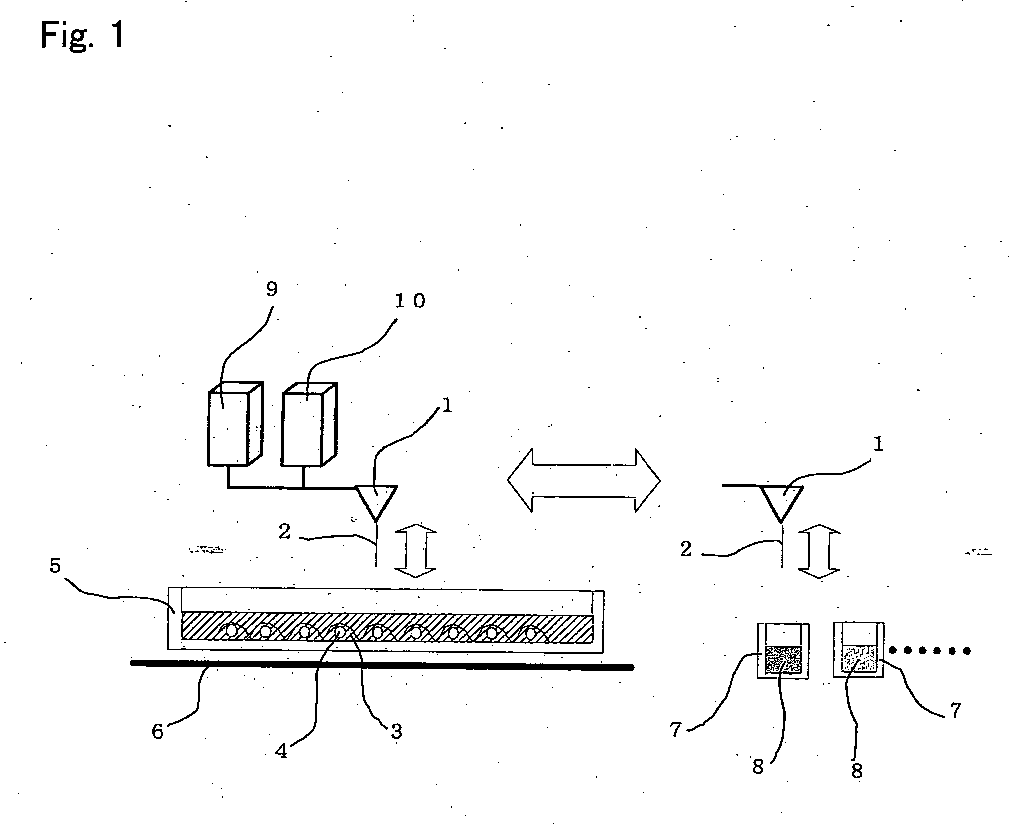
[0033] Preferably, a microinjection device which comprises a washing tank for eliminating the physiologically active substance attached to the surface of the above-described needle, is provided.
[0035] Preferably, the time required for application of a voltage to the above-described needle is shorter than the time at which the above-described needle stays in a cell.

Problems solved by technology

However, it has been difficult to inject a substance into a specific cell.
However, since a hollow glass capillary has been used as a needle to be injected, there has been a certain limit regarding reduction in the external diameter thereof.
Thus, these conventional methods have been problematic in that a cell bursts or suffers fatal damage when a needle is injected therein, or in that operations become complicated.
However, a method for successively recording the change in such a cell over time by actively introducing a gene therein has not yet been disclosed.
In addition, the aforementioned method has been problematic in that the method comprises a complicated step of modifying the surface of a needle with a substance that is allowed to specifically bind to biomolecules.

Method used


Image

  • Microinjection method and device
  • Microinjection method and device
  • Microinjection method and device

Examples


example 1

[0099] Using the device shown in FIG. 1, DNA was introduced into nerve cells that were cultured in a petri dish for culture.

[0100] As nerve cells, PC12. cells (nervous system clone cells isolated from rat adrenal medulla pheochromocytoma) were used. As a medium, DMEM (Dulbecco's Modified Eagle Medium) containing 10% fetal bovine serum (FBS) was used. The culturing was carried out at 37° C. in 5% CO2. As DNA, a recombinant expression vector containing NGF receptor gene was used, and 1 μg / ml DNA solution was used.

[0101] The needle used for the device shown in FIG. 1 composed of the carbon nanotube shown in FIG. 11, which had a diameter of 50 nm and a length of 3 μm.

[0102] First, the needle was immersed in the DNA solution, so as to allow DNA to attach to the surface thereof. Thereafter, the needle was inserted into the nucleus of a nerve cell, and then released the DNA therein. After completion of the introduction of the DNA, the nerve cell was continuously cultured. Even 3 days la...


PUM

PropertyMeasurementUnit
Lengthaaaaaaaaaa
Diameteraaaaaaaaaa
Diameteraaaaaaaaaa

Abstract

An object of the present invention is to provide a method for introducing a physiologically active substance such as a gene into a cell, which introduces a physiologically active substance such as any given gene into any given cell in a view under a microscope, while significantly reducing invasiveness to the cell, and a device used for the above method. The present invention provides a method for introducing a physiologically active substance into a cell, which comprises: allowing a physiologically active substance to attach around a needle having a diameter of 500 nm or less, provided that it is able to be inserted into a cell; and inserting the above-described needle into the cell; and a microinjection device for carrying out the aforementioned method.

Description

TECHNICAL FIELD [0001] The present invention relates to a method for introducing a physiologically active substance into cells and a microinjection device used for the above method. BACKGROUND ART [0002] Examples of a technique of introducing gene DNA into cultured cells or the like may include the calcium precipitation method, the lipid transfer method, the viral vector method, electroporation, the gene gun method, and the microinjection method. In the above methods other than the microinjection method, DNA is introduced in cells at a certain probability, and thus it is impossible to introduce DNA into only a specific cell. On the other hand, the microinjection method has been problematic in that since the diameter of the edge of a glass pipette is approximately 1 μm, cells are easily damaged when such a glass pipette is inserted into the cell nucleus thereof. In addition, when different genes are introduced into multiple cells, the same number of pipettes as that of genes should b...

Claims


Application Information

Patent Timeline
IPC IPC(8): C12N15/00C12M1/00C12M3/00
CPCB82Y30/00C12M35/00
Inventor MIYAWAKI, ATSUSHIIMABAYASHI, HIROYUKIKARAKI, SACHIKO
Owner OLYMPUS CORP
Learn More