Detection and characterization of psychoactives using parallel multi-site assays in brain tissue

a psychoactive compound and multi-site assay technology, applied in the field of detection and characterization of psychoactive compounds, can solve the problems of large effort to develop methods and devices, large disagreement regarding the dominant frequency of activity, and many psychoactive compounds have a relatively small probability of changing the activity of single neurons or even small groups of neurons

Inactive Publication Date: 2007-04-26
PANASONIC CORP +1
View PDF0 Cites 4 Cited by
  • Summary
  • Abstract
  • Description
  • Claims
  • Application Information

AI Technical Summary

Benefits of technology

[0011] It is also desirable to use a multi-electrode dish (“MED”) to measure individual oscillations or network level baselines and network level responses so that a number of different active or less active sites on the neuronal sample may be simultaneously or sequentially sampled. Use of the MED permits measurement and calculation of spatial relationships; both measured and calculated, amongst the values of the neural oscillations. The multi-electrode nature of the MED also enables the determination and characterization of region-specific effects within the given in vitro neuronal sample.

Problems solved by technology
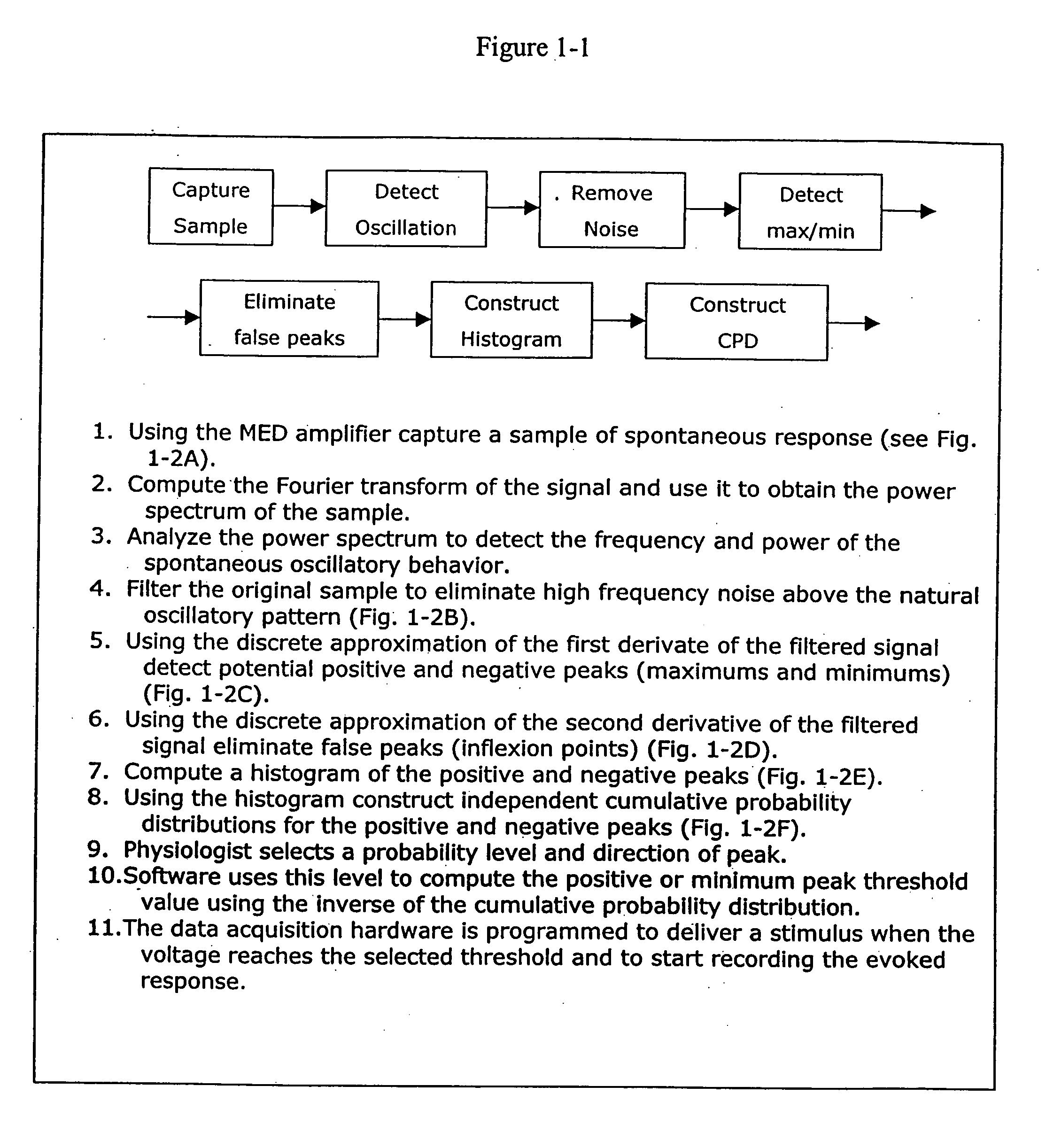
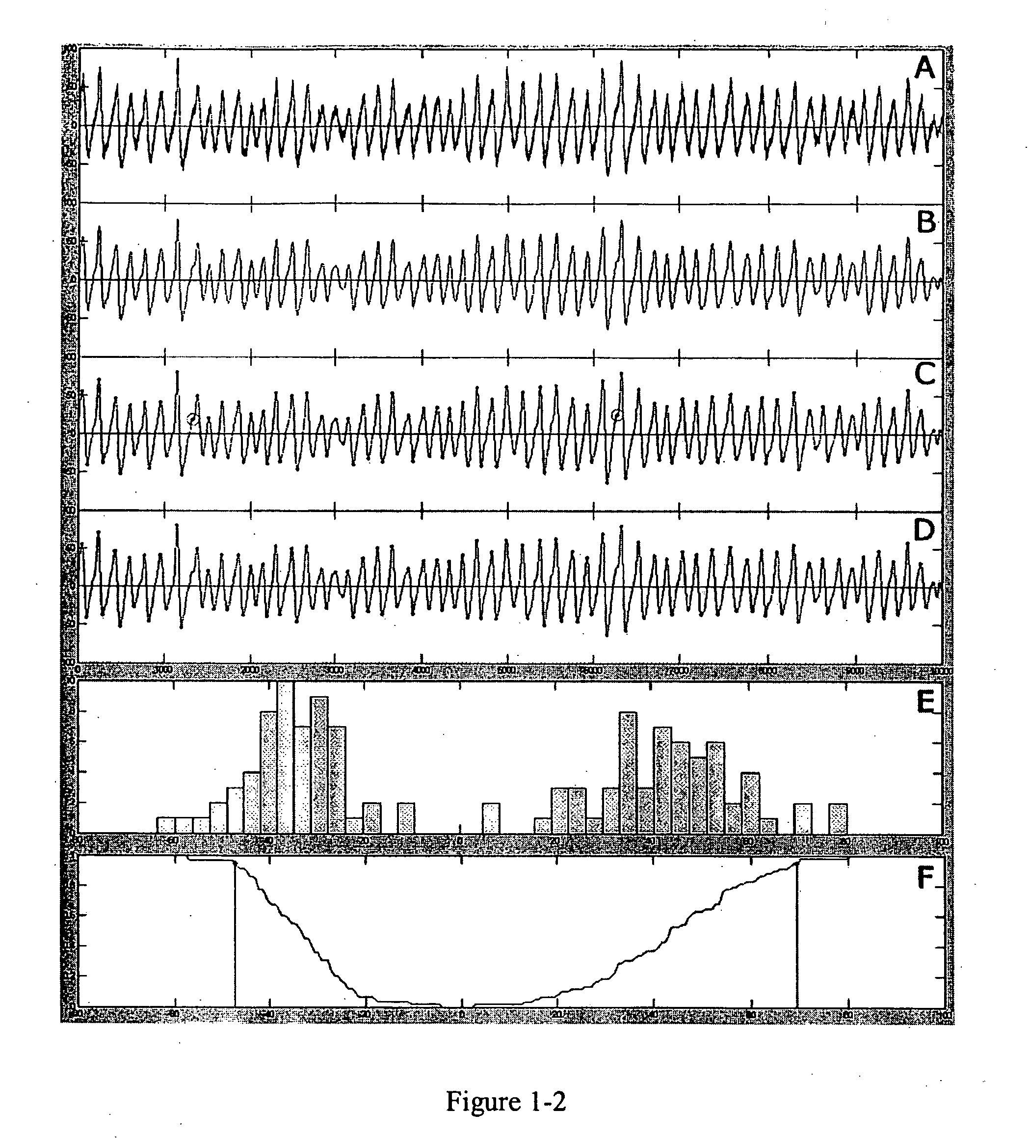
While several studies have shown that infusion of cholinergic agonists into hippocampal slices causes the near immediate appearance of rhythmic oscillations, there is disagreement regarding the dominant frequency of the activity.
Consequently, there has been considerable effort to develop methods and devices for the characterization and detection of psychoactive compounds using carbachol-induced rhythmic oscillations.
A problem encountered in such efforts has been that at low concentrations, many psychoactive compounds have a relatively small probability of changing the activity of single neurons or even small groups of neurons (e.g., currents, firing rate).
Although there may be compound-induced changes at such levels of observation, the changes may be too weak to detect and / or such changes may occur with low probability.
However, many in vivo or in vitro models lack some or all of these important features.

Method used


Image

  • Detection and characterization of psychoactives using parallel multi-site assays in brain tissue
  • Detection and characterization of psychoactives using parallel multi-site assays in brain tissue
  • Detection and characterization of psychoactives using parallel multi-site assays in brain tissue

Examples


example 1

Preparation of Multi-Electrode Array

[0071] Procedures for the preparation of the recently introduced Multi-Electrode Dish (Panasonic: MED probe) are described by Oka et al., J Neurosci Methods 93: 61-67 (1999). The device has an array of 64 planar microelectrodes, each having a size of 50×50 μm, arranged in an 8 by 8 pattern. Probes used in the experiments described below were medium arrays with 300 μm interelectrode spacing (Panasonic: MED-P530AP).

[0072] For sufficient adhesion of the slice to the probe, the surface of the MED probe was treated with 0.1% polyethylenimine (Sigma: P-3143) in 25 mM borate buffer, pH 8.4, for 8 hours at room temperature. The probe surface was rinsed 3 times with sterile distilled water. The probe (chamber) was then filled with DMEM / F-12 mixed medium, containing 10% fetal bovine serum (GIBCO: 16141-079) and 10% horse serum (GIBCO: 16050-122), for at least 1 hour at 37° C. DMEM / F-12 mixed medium is a 1:1 mixture of Dulbecco's Modified Eagle's Medium an...

example 2

Preparation of Hippocampal Slices

[0073] A 17-25 day old Sprague-Dawley rat was sacrificed by decapitation after anesthesia using halothane (2-Bromo-2chloro-1,1,1-trifluoroethane, Sigma: B4388), and the whole brain was removed. The brain was immediately soaked in ice-cold, oxygenated preparation buffer of the following composition (in mM): 124 NaCl, 26 NaHCO3, 10 glucose, 3 KCl, 1.25 NaH2PO4, 1.5 CaCl2, 0.5 MgSO4, for approximately 2 minutes. Appropriate portions of the brain were trimmed and placed on the ice-cold stage of a vibrating tissue slicer (Leica: VT-1000S). The stage was immediately filled with both oxygenated and frozen preparation buffers. The thickness of each tissue slice was 350 μm. Each slice was gently taken off the blade using a blunt-end pipette, and immediately soaked in oxygenated preparation buffer for at least 1 hour at room temperature. A slice was then placed on the center of the MED probe, positioned to cover the 8×8 array. After positioning the slice, the...

example 3

Electrophysiological Recording

[0074] During electrophysiological recording, the slices on the MED probe were placed in a small CO2 incubator (Yamato: model IC400) at 32° C. The slices remained on the MED probe, which was attached to a recording interface, and a moisturized 95% O2 and 5% CO2 gas mixture was blown from above. In this condition, responses were recorded for more than 2 hours.

[0075] Carbachol was obtained from Sigma. A carbachol solution was applied to the neuronal samples at known concentrations of either 25 or 50 μM. The lower concentration was used in all except two cases. In these two cases, the concentration was increased to 50 μM after 25 μM was found to be insufficient to induce powerful oscillations. Carbachol solutions were prepared daily from frozen aliquots.

[0076] Spontaneous and evoked field potentials at all 64 sites were recorded simultaneously with the multi-channel recording system (Panasonic: MED64 system) at a 20 kHz sampling rate. In the case of the...


PUM

PropertyMeasurementUnit
timeaaaaaaaaaa
timeaaaaaaaaaa
timeaaaaaaaaaa

Abstract

This invention relates to methods and devices for the detection and characterization of psychoactive compounds by analyzing alterations of network level physiological characteristics before and after the introduction of a candidate sample onto an in vitro neuronal tissue sample. The invention further provides a software package that enables an operator to deliver a timed electrical pulse to neuronal samples at a specific point in their spontaneous or induced oscillations. Such temporal stimulations trigger unexpected and useful network level physiological responses.

Description

FIELD OF THE INVENTION [0001] The present invention relates to a method and device for the detection and characterization of psychoactive compounds. Specifically, the detection and characterization of psychoactive compounds using network level responses in neuronal tissue samples is described. BACKGROUND OF THE INVENTION [0002] The great majority of synapses in the hippocampus arise from associational and cortical afferents that use glutamate as a transmitter. As with other telencephalic areas, the hippocampus also receives significant projections from several subcortical structures that utilize an array of transmitters other than glutamate. The largest and best studied of the subcortical projections to the hippocampus is the cholinergic input from the medial septum / diagonal bands. These afferents generate the 4-7 Hz theta rhythm by mechanisms that are now fairly well understood (Vertes et al., Neuroscience 81: 893-926 (1997)). Much less is known about how the cholinergic inputs mod...

Claims


Application Information

Patent Timeline
Patent Type & Authority Applications(United States)
IPC IPC(8): C12Q1/00G06F19/00G01N33/483C12M1/34G01N27/30G01N27/416G01N33/15G01N33/48G01N33/487G01N33/50G01N37/00
CPCG01N33/5058
Inventor LYNCH, GARYCOLGIN, LAURA L.SAAVEDRA, RAFAEL H.
Owner PANASONIC CORP
Why Eureka
Try Eureka