System and method for minimally invasive disease therapy

a disease therapy and minimally invasive technology, applied in the field of surgical system and method for removing and treating diseased tissue, can solve the problems of frequent visits, undesirable side effects, and cancerous cells remaining near the resection si

Active Publication Date: 2007-05-31
SUROS SURGICAL SYST
View PDF55 Cites 51 Cited by
  • Summary
  • Abstract
  • Description
  • Claims
  • Application Information

AI Technical Summary

Problems solved by technology

However, unless a large portion of healthy tissue is also resected, a possibility exists that some cancerous cells remain near the resection site.
However, broad-based radiation therapy requires multiple exposures to high doses of radiation.
Such exposure resul...

Method used


Image

  • System and method for minimally invasive disease therapy
  • System and method for minimally invasive disease therapy
  • System and method for minimally invasive disease therapy

Examples


embodiment 400

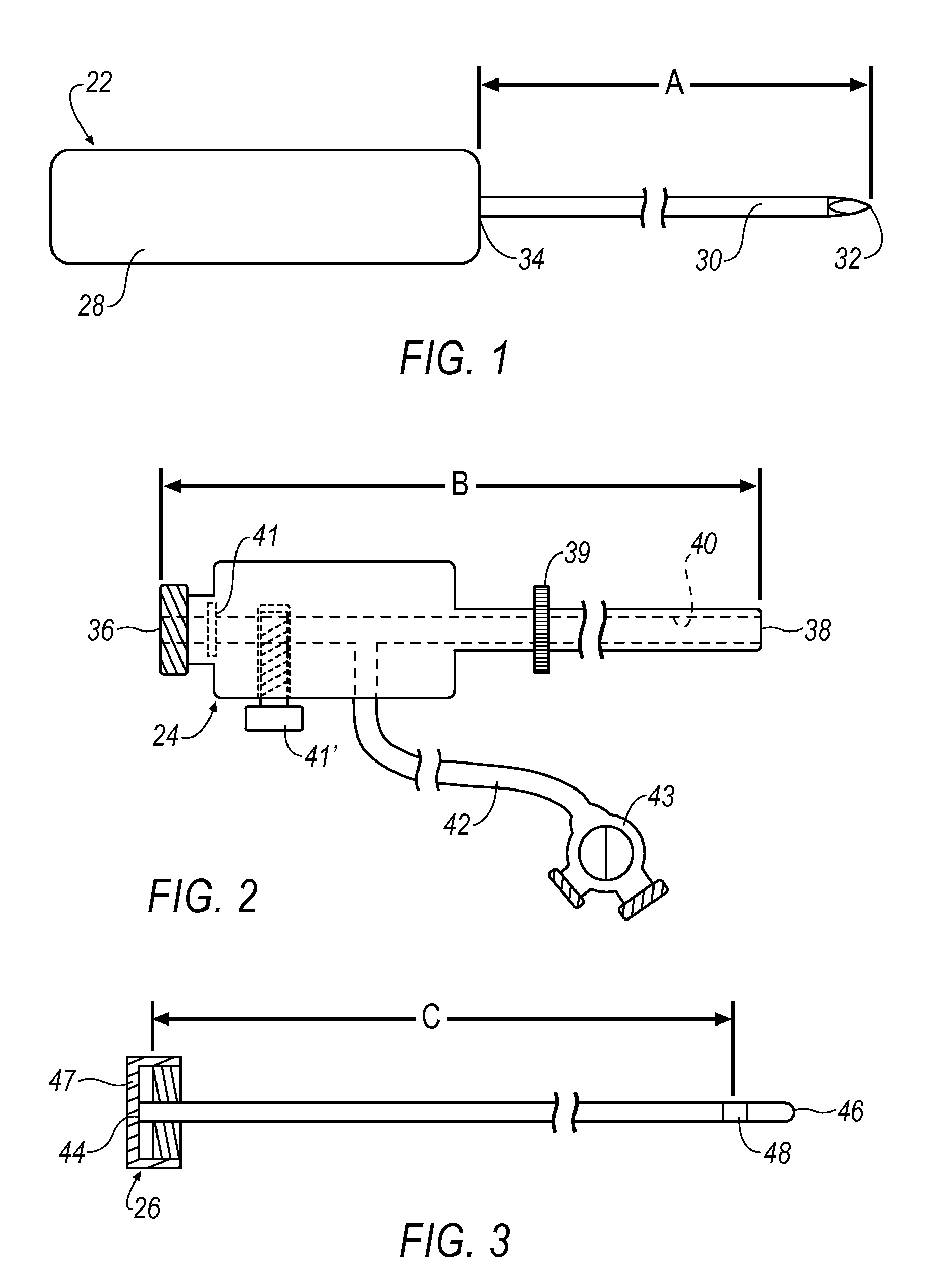
[0096]FIG. 22 shows a haemostatic agent embodiment 400 that introduces a haemostatic agent (represented by arrow(s) H) to target site 182 through valve 43 and fluid conduit 43. A syringe 401 (or other suitable applicator) is attached via a connecting tube 402 to fluid conduit 42. Haemostatic agent H is transported along a lumen 404 formed between outer cannula 56 and inner lumen 40 of outer cannula 24. Check valve 41 functions to prevent haemostatic agent H from exiting outer cannula 24 at proximal end 36. Haemostatic agent H is then deployed at 406 to target site 182 topically, e.g., haemostatic agent H is applied to a surface of target site 182. Once haemostatic agent H is deployed at 406, a haemostatic region 408 is formed that reduces, or in some cases, prevents bleeding into target site 182. Alternatively, haemostatic agent H may be introduced to target site 182 through an introducer, a side port, through a lumen, or through a resection device or treatment device itself.

[0097] ...

embodiment 420

[0100]FIG. 23 shows an alternative haemostatic agent embodiment 420 introducing a haemostatic agent H to target site 182 through proximal end 36 and valve 41. In this embodiment, handpiece 54 is removed from outer cannula 24 and thus, inner lumen 40 is unrestricted (See FIG. 2). Here, syringe 401 is inserted through proximal end 36 and valve 41 such that haemostatic agent H is deployed directly down inner lumen 40 along path 424 (See also FIG. 2). When reaching distal end 38 of outer cannula 24, haemostatic agent H is deployed at 426 to target site 182. In so doing, haemostatic region 408 is formed on an inner wall of target site 182. Depending upon the type and / or form of haemostatic agent H used, selection of a haemostatic deployment embodiment may be selected. For example, a liquid haemostatic agent H may perform well using either the embodiment of FIG. 22 or FIG. 23. However, a dry haemostatic agent H (e.g., a powder) may have improved deployment performance using the embodiment...


PUM


Abstract

A system for treating a lesion site of a patient is disclosed. The system includes a cannula having a lumen, a conduit in communication with said lumen, an introducer stylet removably disposed within said cannula, a resecting device selectively insertable within said cannula, and an adjuvant treatment device selectively insertable within said cannula.

Description

RELATED APPLICATIONS [0001] The present application is a continuation in part application of U.S. patent application Ser. No. 11 / 237,110 filed on Sep. 28, 2005, entitled “System and Method for Minimally Invasive Disease Therapy,” the contents of which are incorporated herein by reference in its entirety.FIELD OF THE INVENTION [0002] The present invention relates generally to a surgical system and method for removing and treating diseased tissue and more particularly to a minimally invasive system and method for removing diseased tissue and creating a margin around an excised area. BACKGROUND Description of the Related Art [0003] Surgical cancer treatments have advanced to two primary stages. A first stage removes the cancerous tissue by resecting the tissue from the body. The goal of the first stage is to remove all cancerous cells from a target area. However, unless a large portion of healthy tissue is also resected, a possibility exists that some cancerous cells remain near the re...

Claims


Application Information

Patent Timeline
IPC IPC(8): A61B17/20
CPCA61B10/0041A61B10/0275A61B10/0283A61B17/32002A61B17/3468A61B17/3478A61B19/20A61B2017/3411A61B2018/0262A61B2019/205A61B2019/304A61B2019/5408A61B2019/5454A61N5/0601A61N5/062A61N5/1015A61B90/10A61B90/17A61B2090/034A61B2090/3908A61B2090/3954
Inventor MARK, JOSEPH L.
Owner SUROS SURGICAL SYST
Why Eureka
Try Eureka