Method and system of computer-aided quantitative and qualitative analysis of medical images

a medical image and quantitative and qualitative technology, applied in image enhancement, instruments, applications, etc., can solve the problems of blood pooling around the tumor, only limited specificity level, and number of unnecessary biopsies, and achieve optimal discrimination of suspicious abnormalities

Active Publication Date: 2007-06-14
SALIENT IMAGING +1
View PDF18 Cites 119 Cited by
  • Summary
  • Abstract
  • Description
  • Claims
  • Application Information

AI Technical Summary

Benefits of technology

[0015] The invention combines both quantitative and qualitative features to achieve an optimal discrimination of suspicious abnormalities, such as imaged breast lesions. Images and data from multiple modalities are processed and analyzed to extract the quantitative and qualitative information. Quantitative information can include kinetics information and biochemical information. Kinetics features can be extracted from a time sequence of image data, such as MRI image data. Biochemical information can be extracted from a spectroscopic analysis of MRS data. Morphological features can be extracted from MRI images, ultrasound images, x-ray images, or images of other modalities. A computer application program is provided for extracting quantitative and qualitative features from medical images and data and for combining results from quantitative and qualitative analysis to produce a consolidated result. The analysis of time course kinetics can be performed before or after the evaluation of the lesions morphology in post-contrast images. Optionally, the results from the first performed analysis are evaluated prior to performing the next analysis. In those cases, if results from the first performed analysis (for example, kinetics analysis) are clearly suggestive, the next analysis (for example, morphology analysis) is not performed. If the results from the analysis of one mode (for example, kinetics) are indeterminate or suggest benign lesion, a further analysis (for example, morphology) is performed.

Problems solved by technology

These new vessels proliferate in a disorganized manner and are poor quality, thus making them leaky and causing blood to pool around the tumor.
However, although the contrast-enhanced MRI method has achieved high levels of sensitivity (94%-100%), it provides only limited specificity levels (40%-95%).
The low specificity levels are result of not only malignant lesions enhancement but also enhancement of the benign lesions, causing a number of unnecessary biopsies.
Thus, the presence of enhancement alone cannot be used to differentiate benign from malignant lesions.
The belief is that the presence of contrast enhancement alone cannot be used to differentiate benign from malignant lesion.
However, correlating spectroscopic data with a scanned image is generally a difficult task in clinical environments.
The forgoing creates challenges for developing a system and method of analyzing medical images for discriminating between malignant and benign lesions, suitable for clinical needs.

Method used


Image

  • Method and system of computer-aided quantitative and qualitative analysis of medical images
  • Method and system of computer-aided quantitative and qualitative analysis of medical images
  • Method and system of computer-aided quantitative and qualitative analysis of medical images

Examples


Embodiment Construction

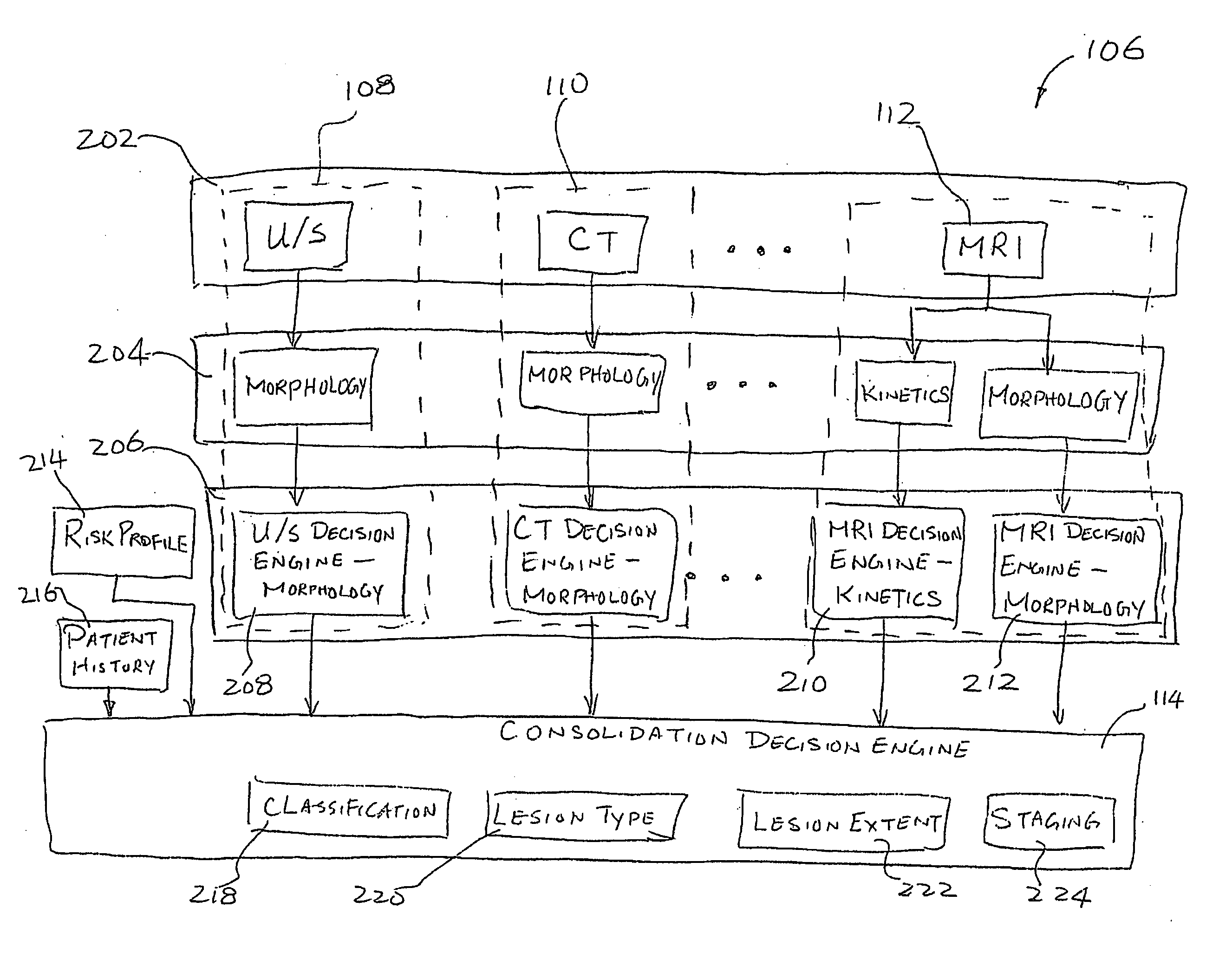
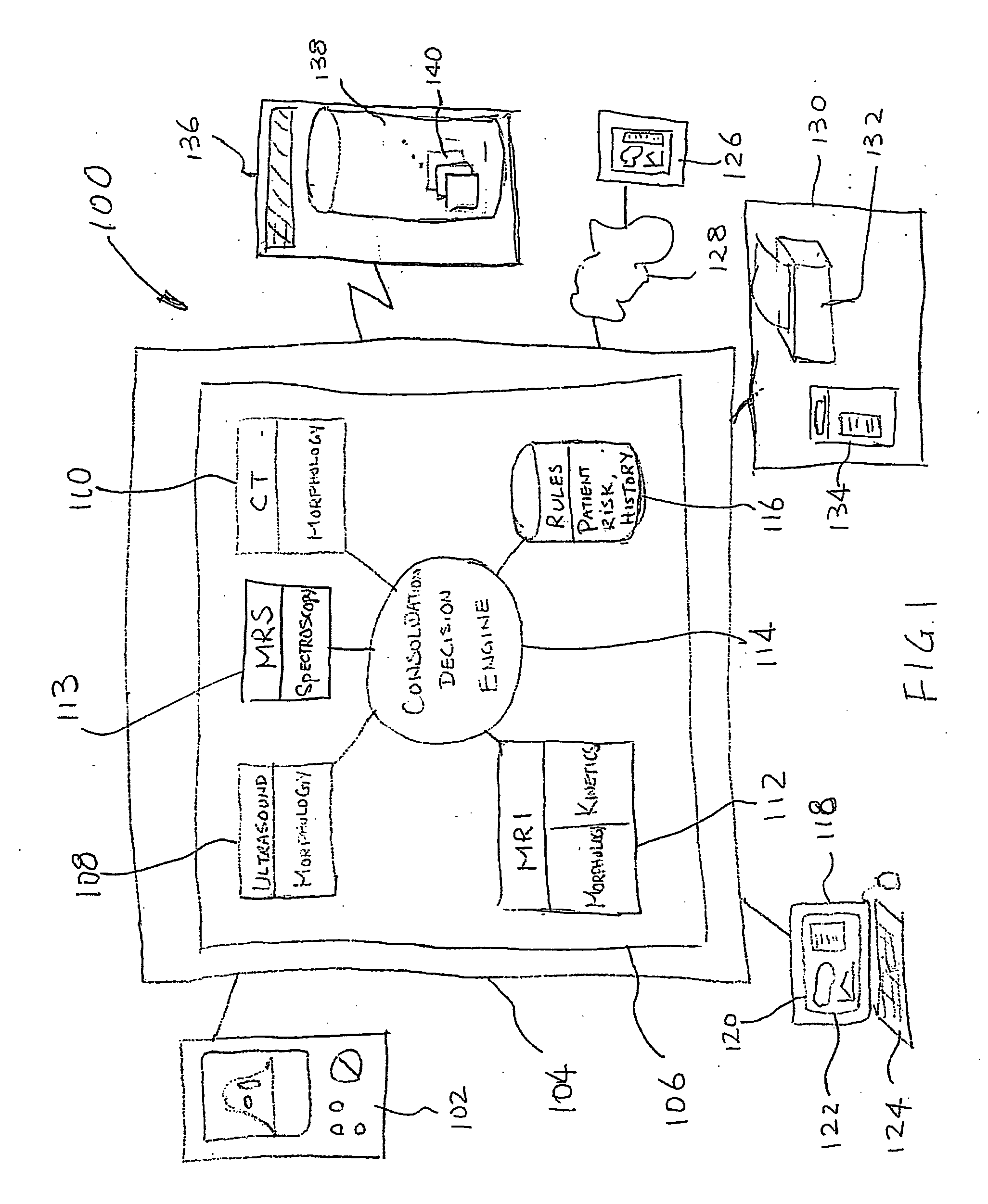
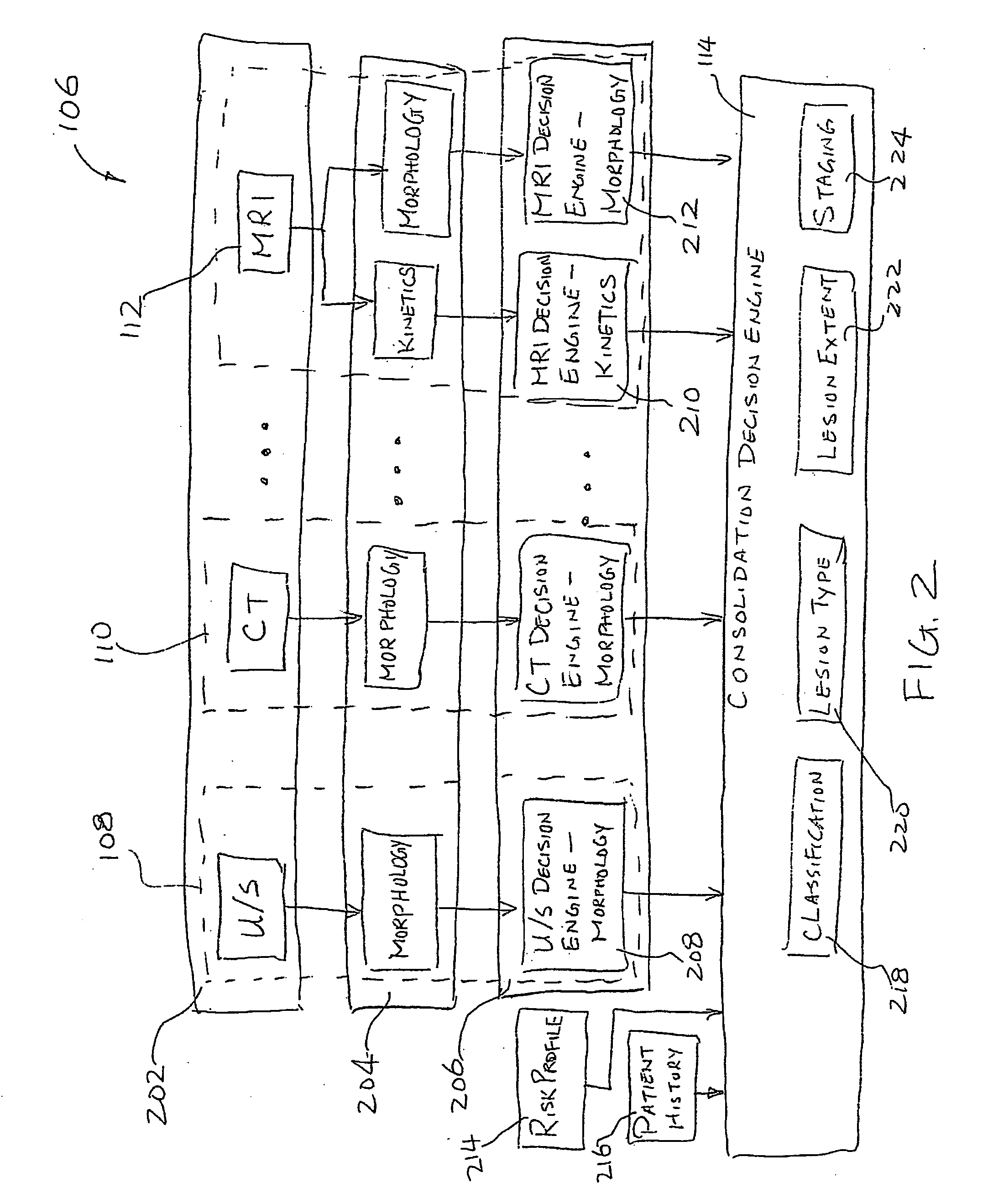
[0040] The invention relates generally to the field of computer-aided analysis of medical images and detection of suspicious abnormalities. In particular, the invention relates to a method and system for processing medical images obtained from multiple modalities, including analysis of kinetics as well as morphological features.

[0041] The invention combines data from multiple modalities, including kinetics (quantitative), morphological (qualitative) and biochemical (quantitative) information to achieve an optimal discrimination of imaged suspicious abnormalities, such as imaged breast lesions. Morphological features of a lesion are generally those associated with size, shape, signal distribution within a mass, or border characteristics of the lesion. They include features such as whether a lesion is a mass having a round, oval or lobular shape, a mass with smooth, irregular or spiculated borders, or a mass having homogeneous, peripheral or ductal enhancement. Morphological features...


PUM


Abstract

A system and method of computer aided analysis of medical images and detection of malignant lesions is described. Medical images obtained from multiple modalities are analyzed. Morphological features as well as temporal, i.e., kinetics features, are combined to compute a consolidated assessment of a possible lesion detected in the medical images. The system includes at least one kinetics module, which is capable of extracting kinetics features from a time sequence of MRI images or MRS data taken after administering a contrast enhancement agent to a patient. The consolidated assessment is presented to a user for confirmation or modification.

Description

CROSS-REFERENCE TO RELATED APPLICATIONS [0001] The present application claims priority from U.S. Provisional Application No. 60 / 738,999 filed on Nov. 23, 2005, the content of which is hereby incorporated by reference.FIELD OF THE INVENTION [0002] The invention relates generally to the field of computer-aided analysis of medical images and detection of suspicious abnormalities. In particular, the invention relates to a method and system for processing medical images obtained from multiple modalities, including analysis of kinetics as well as morphological features and automated detection of abnormalities in and analysis of medical images from multiple modalities. BACKGROUND OF INVENTION [0003] Magnetic resonance imaging (MRI) is emerging as a powerful tool for the imaging of breast abnormalities. In general, MRI provides a better characterization of the breast lesions than conventional imaging modalities due to rich soft-tissue contrast, thin-section and multiplanar capabilities. [00...

Claims


Application Information

Patent Timeline
Patent Type & Authority Applications(United States)
IPC IPC(8): G06K9/00G06V10/764
CPCA61B5/055A61B6/03A61B8/08G01R33/5601G01R33/5608G01R33/56366G06K9/00G06K2209/053G06T5/50G06T7/0012G06T2207/10016G06T2207/20036G06T2207/30068A61B6/5247G06V2201/032G06V10/764A61B6/00G01R33/465G06F18/00
Inventor COLLINS, JEFFREYLACHMANN, FREDERICSAGHATELYAN, KARENSTAPLETON, SANDRA
Owner SALIENT IMAGING
Why Eureka
Try Eureka