

Protein markers for esophageal cancer
a technology of esophageal cancer and protein markers, applied in the field of proteins, can solve the problems of poor survival after diagnosis and far advanced stag
- Summary
- Abstract
- Description
- Claims
- Application Information
AI Technical Summary
Benefits of technology
Problems solved by technology
Method used
Image

Examples
example 1
Carrier Ampholyte (CA) Based 2-D Gels of Esophageal Cancer.
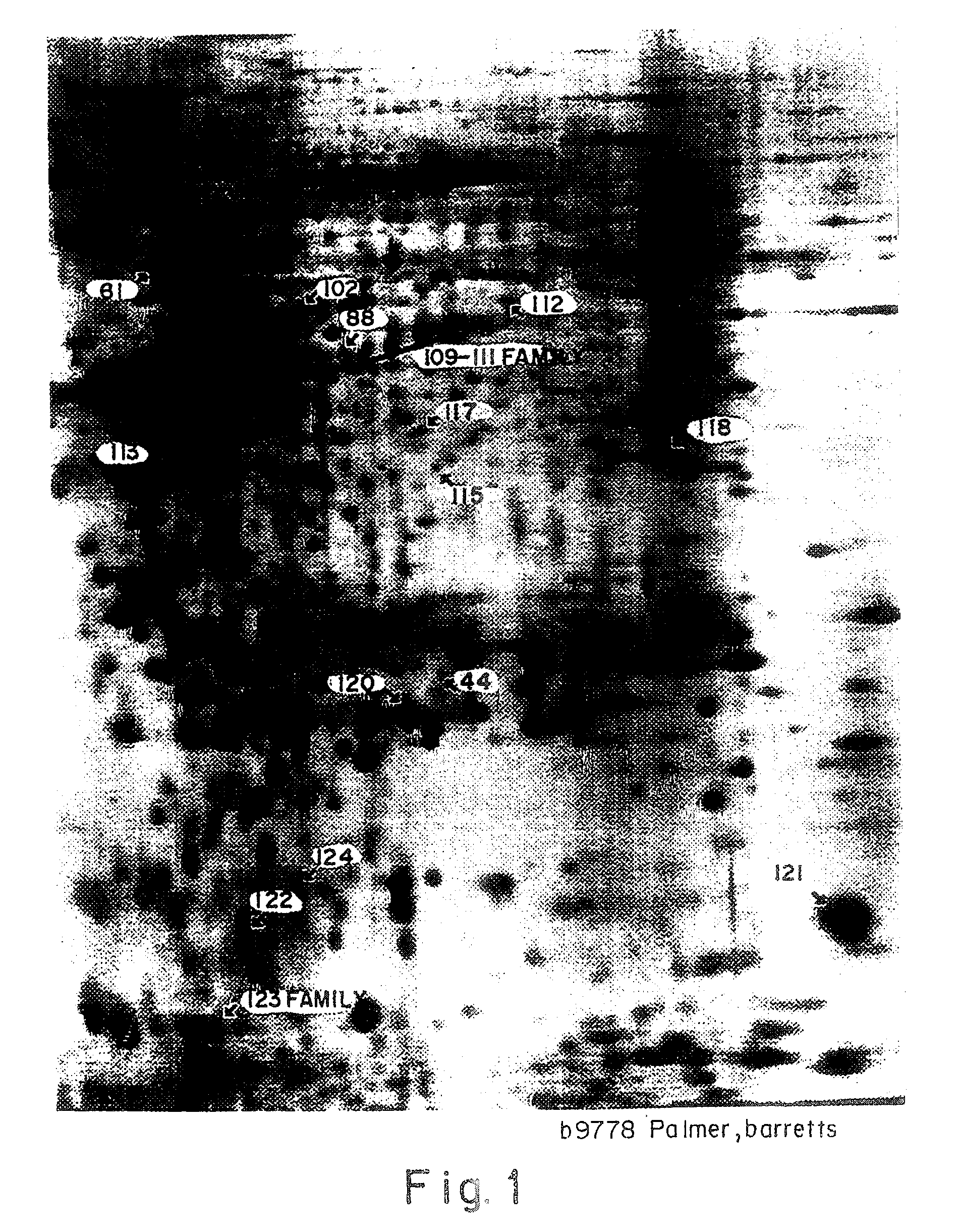
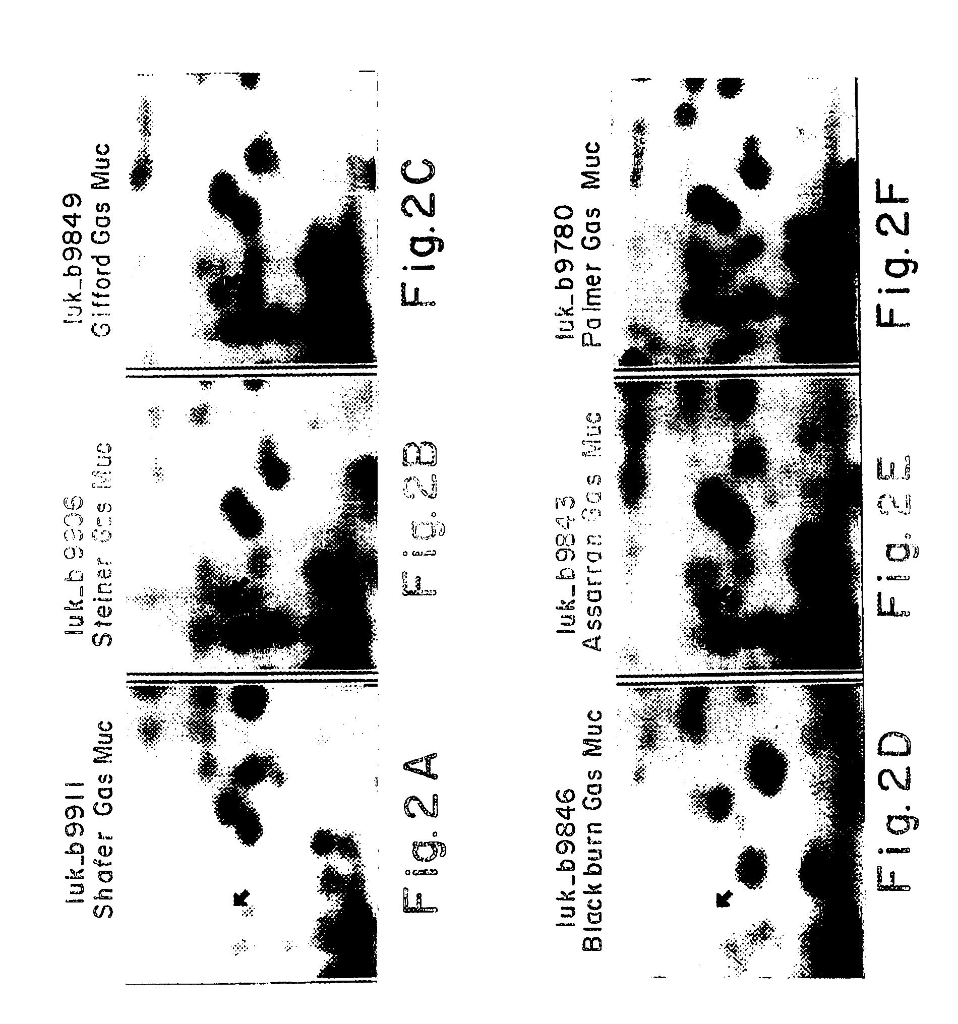
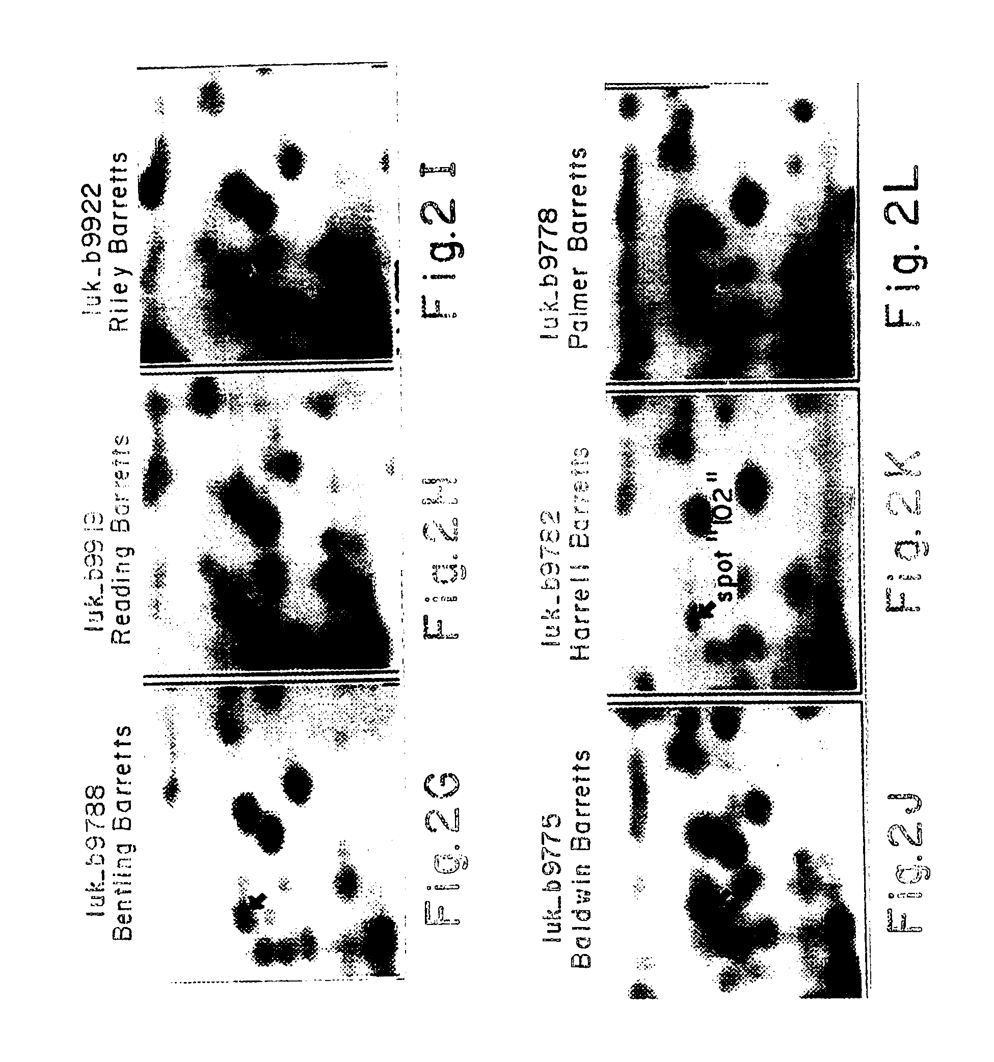
[0035] Tissue from 27 patients was obtained for 2-D analysis from tumor tissue and normal esophagus. For patients in whom the tumor occurred in the context of Barrett's mucosa, Barrett's tissue was also obtained. (An interesting pathological feature of Barrett's mucosa is that it resembles gastrointestinal type of tissue rather than esophageal tissue, where it is found. The lower end of the esophagus resembles gastric mucosa rather than true esophageal tissue so that tumors that arise in that location need to be compared to normal esophagus as well as gastric tissue as control.) For tumors that occurred at the lower end of the esophagus, normal gastric mucosa was also obtained. All specimens were obtained at the time of initial surgery. Therefore, for every patient two control tissues were obtained, in addition to the cancer: normal esophagus and Barrett's for half of the patients, and normal esophagus and stomach for the ...
example 2
Immobilized pH Gradient (IPG) 2-D Gels of Esophageal Cancer
[0040] In addition to generating 2-D patterns that were carrier ampholyte-based, a second set of patterns using immobilized pH gradients were generated as a complementary set.
[0041] Samples were prepared as for the CA gels discussed in Example 1. For first dimension separation an immobilized pH gradient covering the separation range of pH 4-10. The second dimension is the same as for the CA gels of Example 1.
[0042] IPG gels are prepared using derivatives of acrylamide having carboxyl or tertiary amino groups with specific pK values. A linear pH gradient is prepared from a dense, acidic solution and a light, basic solution using a two-chamber microgradient former. The pH gradient is stabilized during polymerization of the Immobiline-acryl-amide-bisacrylamide matrix by a co-linear gradient of glycerol. Formulations of buffering Immobiline mixtures with titrating Immobiline for the pH limit solutions for narrow pH gradients...
example 3
Computer Assisted Analysis of 2-D Gels
[0044] Each gel was scanned in a 1024×1024 pixel format, where each pixel can have one of 256 possible values representing different degrees of intensity. Spot lists for study images are matched to spot lists of master images so that the result is a hierarchy of matched protein spots. The purpose of the matching is to link the same polypeptide spot through the hierarchy to allow assessment of its present, quantitative variation and specificity, as described in Strahler et al, 1990 (J. Clin. Invest. Vol. 85, pp 200-207). For comparison, between gels, of the amount of individual proteins, an adjustment process is utilized. The integrated intensity of detected polypeptides, measured in units of optical density per square millimeter, is adjusted relative to the intensity of reference polypeptides that are ubiquitously expressed. The adjustment is made to compensate for any variation between gels due to protein loading or staining.
PUM
| Property | Measurement | Unit |
|---|---|---|
| molecular weight | aaaaa | aaaaa |
| time | aaaaa | aaaaa |
| pH | aaaaa | aaaaa |
Abstract
Description
Claims
Application Information