Medical imaging diagnostic method

a diagnostic method and imaging imaging technology, applied in the field of medical imaging diagnosis device and medical imaging diagnosis method, can solve problems such as inability to accurately measure the image, and achieve the effect of improving the accuracy of measuremen

Inactive Publication Date: 2007-06-28
HITACHI MEDICAL CORP
View PDF3 Cites 17 Cited by
  • Summary
  • Abstract
  • Description
  • Claims
  • Application Information

AI Technical Summary

Benefits of technology

[0029] It means that in color Doppler images, the pixels equivalent of the lumen where blood flows is displayed in color, and the pixels equivalent of the lumen where there is no blood flow is displayed in black and white. Therefore, the pixel coordinate equivalent of the boundary of the lumen and the tunica intima can be detected by determining the color information of the color Doppler image with respect to each pixel. As a result, the boundary of the lumen and the tunica intima (the inner wall of the tunica intima) can be detected even when the local maximal point on the lumen side of the brightness distribution does not appear due to the factors such as noise.

Problems solved by technology

Due to the individual variability of objects being examined, there are cases that IMT cannot be measured accurately.

Method used


Image

  • Medical imaging diagnostic method
  • Medical imaging diagnostic method
  • Medical imaging diagnostic method

Examples


first embodiment

The First Embodiment

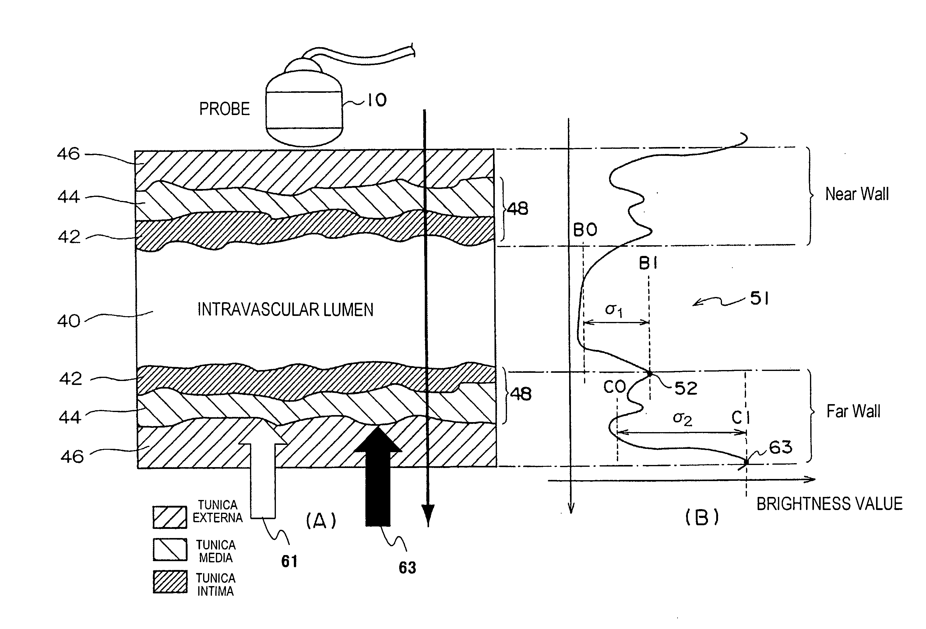
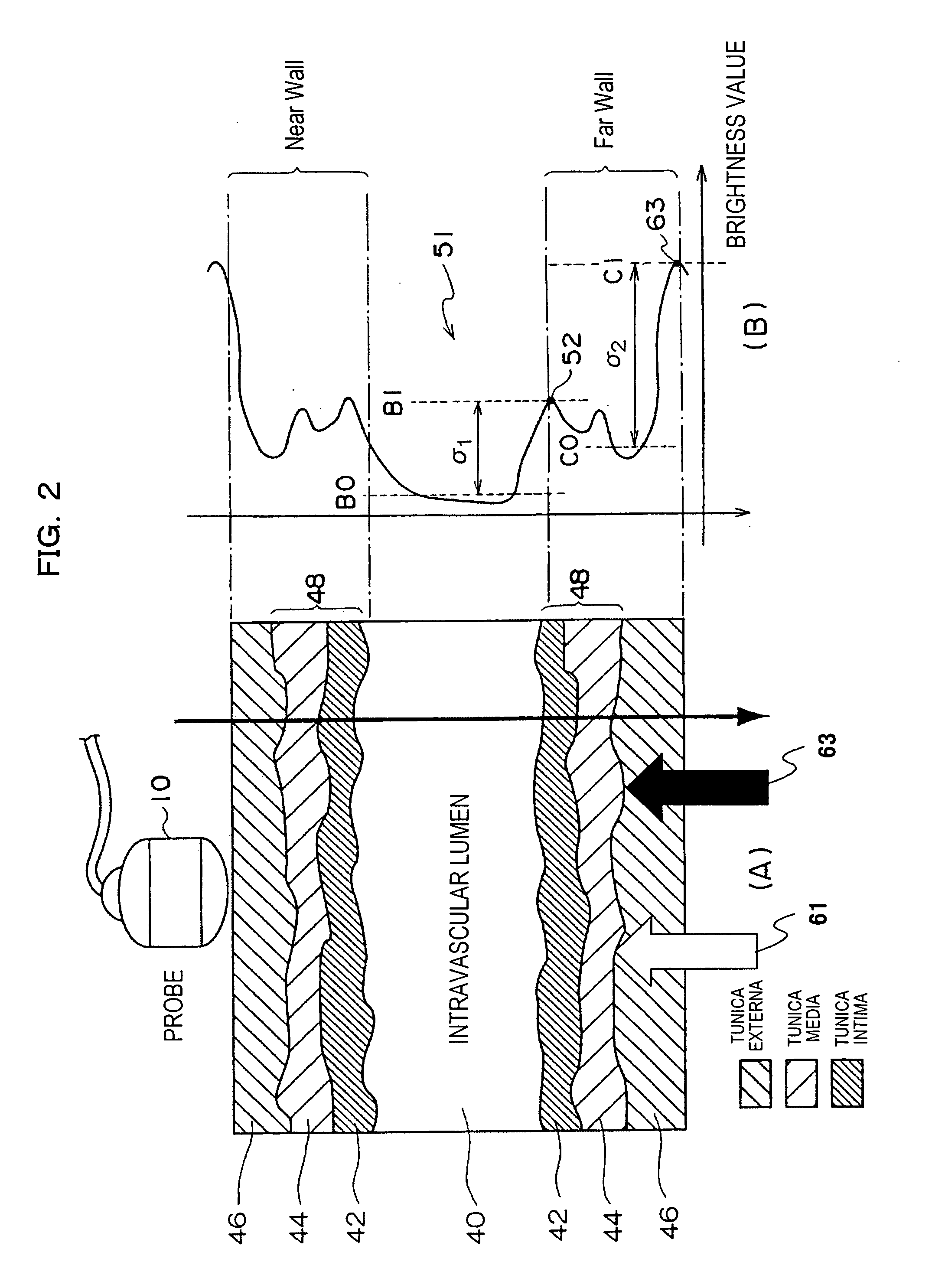
[0044] The first embodiment of the ultrasound diagnostic apparatus to which the present invention is applied as a medical imaging diagnostic apparatus will now be described referring to FIG. 1˜FIG. 6. The present embodiment is an example of measuring IMT (composite thickness of the tunica intima and the tunica media) by applying the region growing method as a region extracting method corresponding to ultrasound images. FIG. 1 is a block diagram of the ultrasound diagnostic apparatus in the present embodiment.

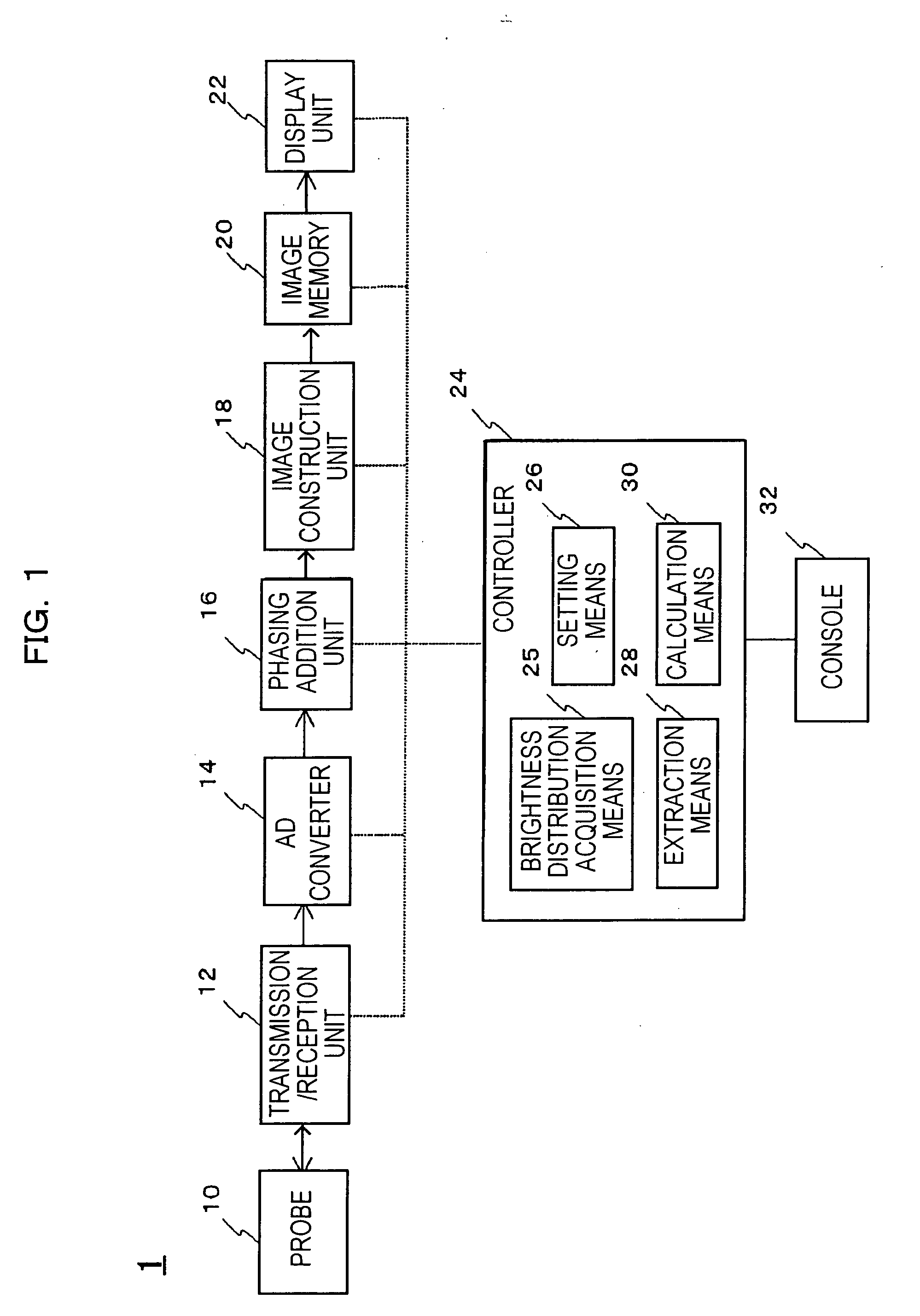
[0045] As shown in FIG. 1, the ultrasound diagnostic apparatus is provided with imaging means for imaging the ultrasonic image with regard to an object (for example, a blood vessel). Imaging means comprises units such as:

[0046] probe 10 for transmitting / receiving ultrasonic waves between the object;

[0047] transmission / reception unit 12 for providing drive signals to probe 10 as well as receiving the transmission of reflected echo signals outputted from pro...

third embodiment

The Third Embodiment

[0081] The third embodiment of the ultrasound apparatus to which the present invention is applied as a medial imaging diagnostic apparatus will now be described referring to FIG. 7. The difference of the present embodiment from the first embodiment is to decrease the error of setting tunica intima SP54 by mistake. Thus the description of the present embodiment will focus on differences from the first embodiment. Places that are mutually corresponding will be described with the same encoding.

[0082] In brightness distribution line 51 of FIG. 2, there are occasions that tunica intima SP54 instead of tunica intima 42 is set by mistake when, for example, a plurality of local maximal points appear on the side of lumen 40 due to factors such as noise. Given this factor, in the present embodiment, a plurality of brightness distribution lines in the vessel diameter direction (depth direction) is obtained over the blood flow direction (lateral direction), and tunica intim...

fourth embodiment

The Fourth Embodiment

[0086] The fourth embodiment of the ultrasound apparatus to which the present invention is applied as a medical imaging diagnostic apparatus will now be described referring to FIG. 8. The difference of the present embodiment from the first embodiment is that the tunica intima SP54 is modified when it is set by error. Therefore, the description of the present embodiment will focus on differences from the first embodiment. Places that are mutually corresponding will be described with the same encoding.

[0087] As shown in FIG. 8, when a plurality of local maximal points 80 and 82 appear on the side of lumen 40 of the brightness distribution line, there are occasions that local maximal point 80 is set by error as tunica intima SP54. By implementing the region extracting method such as region growing method based on the set tunica intima SP54, the region corresponding to tunica intima 42 is extracted. The extracted region cuts across the pre-set range. In other words...


PUM


Abstract

A medical imaging diagnosis apparatus for measuring the composite thickness of the tunica intima (42) and the tunica media (44) of a blood vessel of a subject by acquiring image data on the blood vessel.
In order to improve the accuracy of the IMT measurement of the composite thickness, the medical imaging diagnosis apparatus has extracting means for extracting the tunica intima (42) and the tunica exterma (46) of the blood vessel on the basis of the brightness information of the image data to measure the composite thickness of the tunica intima and the tunica exterma of the vessel in reference to the two extracted regions.

Description

TECHNICAL FIELD [0001] The present invention relates to a medical imaging diagnosis device and medical imaging diagnosis method for performing IMT measurement. BACKGROUND ART [0002] An ultrasound diagnostic apparatus being a kind of medical imaging diagnostic apparatus obtains image data by transmitting / receiving ultrasonic waves between an object being examined via a probe and reconstructing ultrasound images (for example, B-mode images) based on echo signals generated from the object. [0003] In such ultrasound diagnostic apparatus, the measurement of intima media thickness (hereinafter referred to as IMT) of a blood vessel wall is implemented in order to identify arteriosclerosis or cardiovascular diseases at an early stage. A blood vessel wall is formed with a trilaminar structure consisting of tunica intima, tunica media and tunica externa in due order from the lumen side where blood flows. IMT is the sum of the thickness of the tunica intima and the tunica media, in other words...

Claims


Application Information

Patent Timeline
Patent Type & Authority Applications(United States)
IPC IPC(8): A61B5/05A61B8/08
CPCA61B5/02007A61B8/469A61B8/0858
Inventor OSHIKI, MITSUHIROSHINOMURA, RYUICHI
Owner HITACHI MEDICAL CORP
Why Eureka
Try Eureka