Ultrasound imaging system for extracting volume of an object from an ultrasound image and method for the same

an ultrasound image and ultrasound technology, applied in image enhancement, instruments, medical/anatomical pattern recognition, etc., can solve the problems of long time required for extracting prostate contour, difficult to extract prostate contour, and difficult to achieve the effect of reducing noise in the edge image and clear object contour

Inactive Publication Date: 2007-07-19
MEDISON CO LTD
View PDF5 Cites 24 Cited by
  • Summary
  • Abstract
  • Description
  • Claims
  • Application Information

AI Technical Summary

Benefits of technology

[0016] In accordance with the present invention, there are provided an ultrasound imaging system and method for extracting volume data of an object from 3D ultrasound image data by using wavelet transform and SVM. Thus, it is possible to obtain a clear contour of the object while reducing noises in the edge image formed by averaging wavelet-transformed images at respective scales.

Problems solved by technology

Thus, if the prostate is inflamed or enlarged, it can cause various urinary problems.
In such a case, it takes a long time to extract the prostate contour.
On the other hand, the conventional method is disadvantageous in that the user has to manually draw draft contours on all the 2D cross-sectional images obtained from the 3D volume in the snakes algorithm and the contour detection results considerably depend on snakes variables.
However, the time required for extracting the prostate contour can still be long depending on the sizes of input images and the stick-shaped filter.

Method used


Image

  • Ultrasound imaging system for extracting volume of an object from an ultrasound image and method for the same
  • Ultrasound imaging system for extracting volume of an object from an ultrasound image and method for the same
  • Ultrasound imaging system for extracting volume of an object from an ultrasound image and method for the same

Examples


Embodiment Construction

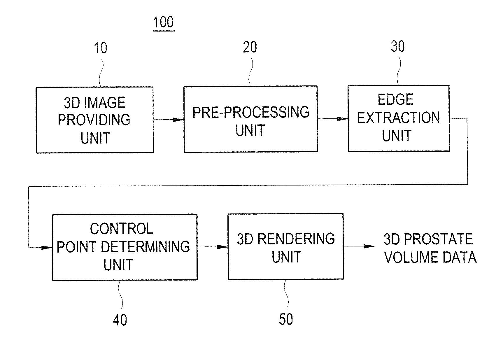
[0033] Hereinafter, an ultrasound imaging system and method for automatically extracting a volume of a target object (for example, a prostate) in accordance with the present invention will be described with reference to the accompanying drawings.

[0034] Referring now to FIG. 1, an ultrasound imaging system 100 for forming volume data of a target object in accordance with one embodiment of the present invention includes a 3D ultrasound image providing unit 10, a pre-processing unit 20, an edge extraction unit 30, a control point determining unit 40 and a 3D rendering unit 50. The 3D image providing unit 10 can be a memory or a probe. The pre-processing unit 20, the edge extraction unit 30, the control point determining unit 40 and the 3D rendering unit 50 can be embodied with one processor. The control point determining unit 40 includes a support vector machine (SVM).

[0035] Here, an edge is a point where the discontinuity of brightness appears. Further, a boundary is a contour of th...


PUM


Abstract

The present invention provides an ultrasound imaging system for forming 3D volume data of a target object, including a three-dimensional (3D) image providing unit for providing a 3D ultrasound image; a pre-processing unit for forming a number of two-dimensional (2D) images from the 3D ultrasound image and normalizing the 2D images to form normalized 2D images; an edge extraction unit for forming wavelet-transformed images of the normalized 2D images at a number of scales, the edge extraction unit further being configured to form edge images by averaging the wavelet-transformed images at a number of scales and threshold the edge images; a control point determining unit for determining control points by using a support vector machine (SVM) based on the normalized 2D images, the wavelet-transformed images and the thresholded edge images; and a rendering unit for forming 3D volume data of the target object by 3D rendering based on the control points.

Description

FIELD OF THE INVENTION [0001] The present invention generally relates to an ultrasound imaging system and a method for processing an ultrasound image, and more particularly to an ultrasound imaging system for automatically extracting volume data of a prostate and a method for the same. BACKGROUND OF THE INVENTION [0002] An ultrasound diagnostic system has become an important and popular diagnostic tool due to its wide range of applications. Specifically, due to its non-invasive and non-destructive nature, the ultrasound diagnostic system has been extensively used in the medical profession. Modem high-performance ultrasound diagnostic systems and techniques are commonly used to produce two or three-dimensional (2D or 3D) diagnostic images of a target object. The ultrasound diagnostic system generally uses a wide bandwidth transducer to transmit and receive ultrasound signals. The ultrasound diagnostic system forms ultrasound images of the internal structures of the target object by e...

Claims


Application Information

Patent Timeline
Patent Type & Authority Applications(United States)
IPC IPC(8): A61B8/00
CPCA61B8/08A61B8/483G06K9/4609G06K9/4671G06K9/6254G06T2207/30081G06T7/0083G06T7/0095G06T2207/10132G06T2207/20064G06K2209/05G06T7/12G06T7/168G06V10/443G06V10/462G06V2201/03G06V10/7788A61B8/00G06F18/41
Inventor KIM, NAM CHULOH, JONG HWANKIM, SANG HYUNKWAK, JONG INAHN, CHI YOUNG
Owner MEDISON CO LTD
Why Eureka
Try Eureka