Surgical Articles for Placing an Implant about a Tubular Tissue Structure and Methods

a tubular tissue and implant technology, applied in the field of surgical instruments, can solve the problems of urethral obstruction, difficulty in voiding, rare complications of sling procedures,

Inactive Publication Date: 2007-08-09
BOSTON SCI SCIMED INC
View PDF34 Cites 18 Cited by
  • Summary
  • Abstract
  • Description
  • Claims
  • Application Information

AI Technical Summary

Benefits of technology

[0018] The repairs of the present invention may be achieved without substantially changing the orientation of the patient's anatomy to avoid, in the case of an incontinence procedure, post-surgical voiding difficulties brought about by the change in the orientation of the natural body passageway (e.g. the urethra or urethral meatus). In some embodiments of the present invention, urinary incontinence may be treated without the need for a significant abdominal incision or, in some embodiments, a small vaginal incision or puncture.
[0031] In another aspect, the present invention comprises a surgical method for placing an implantable article about a tubular tissue structure. The surgical procedure may be for general surgical purposes discussed in greater detail below. The surgical instruments are particularly suitable for treating incontinence. In embodiment, the method comprises the steps of a) inserting a surgical device with an implantable article situated therein into a urethra, b) moving the implantable article from a position within the urethra to a position at least substantially external to the urethra; c) separating the implantable article from the device, and d) removing the surgical device from the urethra leaving the implantable article situated about the urethra. The procedure has, among other advantages, the advantage of treating incontinence without requiring an incision in the vagina or abdomen of the patient.
[0032] In another aspect, the method comprises the steps of: a) providing a surgical device having an immobilizer and a stabilizer, and a implantable article deployment member movable about the immobilizer between a retracted position and an extended position; b) inserting the immobilizer into the urethra and the stabilizer within the vagina; c) moving the implantable article deployment member along a path that is substantially external to the urethra from the retracted to the extended position; d) retracting the implantable article deployment member from the extended to the retracted position leaving the implantable article situated about the urethra; e) separating the implantable article from the device; and f) removing the surgical device from the patient's body. This procedure has the advantage of treating the incontinence without substantially changing the patient's urinary tract orientation to avoid post implantation voiding difficulties

Problems solved by technology

A complication associated with these procedures is voiding difficulties, possibly due to the resultant geometry of the urinary tract.
Bone anchors or screws raise the specter of bone infection, necrosis and other complications, although such complications are rare.
Complications associated with sling procedures are rare, but they include urethral obstruction, infection, development of de novo urge incontinence, bladder perforation, hemorrhage, prolonged urinary retention, and damage to surrounding tissue (e.g. caused by sling erosion).

Method used


Image

  • Surgical Articles for Placing an Implant about a Tubular Tissue Structure and Methods
  • Surgical Articles for Placing an Implant about a Tubular Tissue Structure and Methods
  • Surgical Articles for Placing an Implant about a Tubular Tissue Structure and Methods

Examples


Embodiment Construction

[0070] The following description is meant to be illustrative only and not limiting. Other embodiments of this invention will be apparent to those of ordinary skill in the art in view of this description.

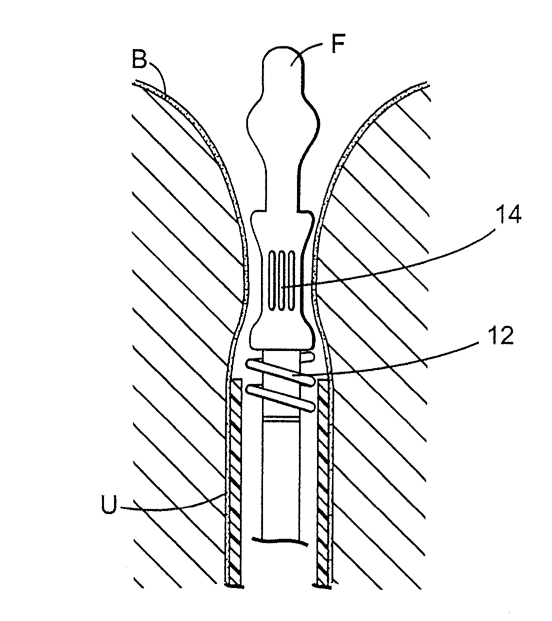
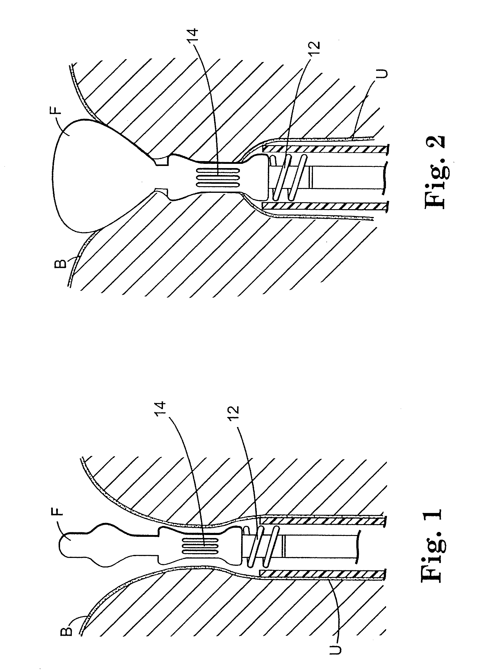
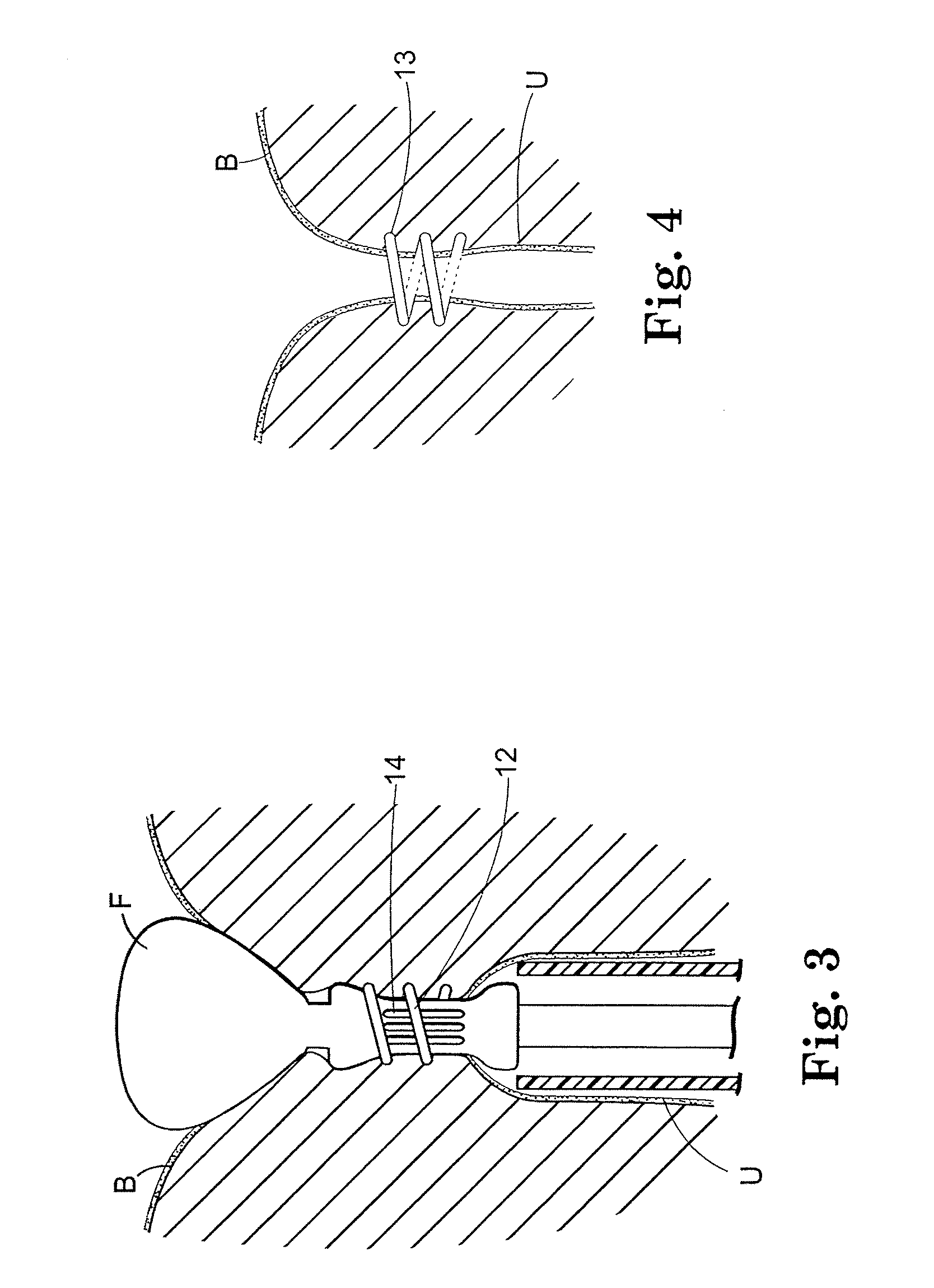
[0071]FIGS. 1 through 4 are schematic illustrations of a surgical device for placing an implantable article about a tubular tissue structure, such as a patient's urethra U. The bladder neck is situated between the patient's bladder B and urethra U.

[0072] The surgical device includes an inflatable member F, a means for collapsing the tubular tissue structure (in this embodiment a vacuum element 14) about an immobilizer (the tubular structure between the vacuum ports), and an implantable article deployment member 12 movable between a retracted position (FIG. 1) and an extended position (FIG. 3).

[0073]FIG. 1 shows the surgical device after it is inserted transurethrally so that portions are deployed within the bladder B and urethra U.

[0074]FIG. 2 shows the inflatable member F inflat...


PUM


Abstract

A minimally invasive surgical instrument for placing an implantable article about a tubular tissue structure is disclosed. The surgical instrument is particularly useful for treating urological disorders such as incontinence. Surgical methods using the novel instrument are also described.

Description

CLAIM FOR PRIORITY [0001] This divisional patent application claims priority to U.S. utility patent application Ser. No. 10 / 233,349, filed Aug. 30, 2002, and entitled “Surgical Articles for Placing an Implant About a Tubular Tissue Structure and Methods,” U.S. provisional patent application No. 60 / 325,870, filed Sep. 28, 2001, and U.S. provisional patent application No. 60 / 316,552, filed Aug. 31, 2001. The identified utility patent application and provisional applications are herein incorporated by reference.BACKGROUND [0002] Examples of surgical instruments for soft tissue repair or manipulation are disclosed in U.S. Pat. Nos. 1,449,087; 3,470,875; 3,763,860; 3,946,740; 4,164,225; 4,923,461; 4,935,027; 5,527,321; 5,431,666; 5,674,230; 5,728,107; 5,730,747, 5,741,279; 5,871,488; 6,056,771 and 6,084,351. [0003] The Capio™ CL Transvaginal Suture Capturing Device, and the Capio™ Suture Capturing Device are available from Boston Scientific, of Natick, Mass. These devices are capable of ...

Claims


Application Information

Patent Timeline
Patent Type & Authority Applications(United States)
IPC IPC(8): A61F2/02A61F2/06A61F2/84
CPCA61B17/0469A61B17/0483A61B2017/00805A61F2002/9517A61B2017/306A61F2/962A61B2017/00827A61F2/0045A61F2/9517
Inventor ANDERSON, KIMBERLY A.WATSCHKE, BRIAN P.MORNINGSTAR, RANDY L.BACHMAN, TIMOTHY A.NEISZ, JOHANN J.
Owner BOSTON SCI SCIMED INC
Why Eureka
Try Eureka