

Method and device for treating microscopic tumors remaining in tissues following surgical resection
- Summary
- Abstract
- Description
- Claims
- Application Information
AI Technical Summary
Benefits of technology
Problems solved by technology
Method used
Image


Examples
example i
Treatment of Apparently Healthy Tissue Surrounding an Open Wound with Electroporation Pulses and / or Anticancer Agent
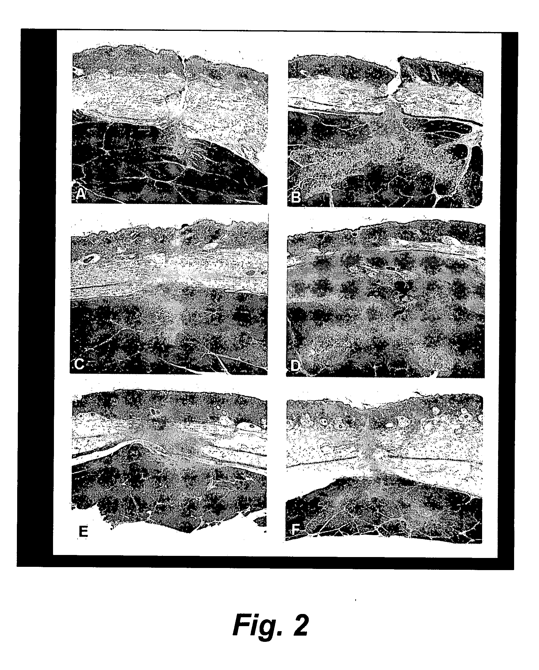
[0040] In order to successfully use electroporation in areas of open wounds comprising for the most part normal tissue, we conducted a series of experiments to show that electroporation of normal tissue, with or without anti-cancer agent, can be performed without significantly affecting the healing processes in cutaneous, subcutaneous and muscle tissues.
[0041] In this experiment eighteen transcutaneous incisions were made into the dorsal muscles in each of eight pig test animals grouped 1-6. In study groups 1-5, longitudinal incisions were made while in group 6 the incisions were transverse to the longitudinal orientation of the muscle fiber. At each incision site, either nothing, normal saline, or Bleomycin in solution was injected such that injections were placed to a 1 cm depth and spaced at 1 cm distances from one another with three injections, one on each side ...
example ii
Hypothetical Treatment Regimen
It is contemplated that surgery using the invention method and device will generally follow a protocol likely to employ the following steps:
[0051] 1. The main tumor mass will be surgically resected as usually practiced by a surgeon skilled in the art. The surgeon will either resect the primary tumor mass only or will also resect a surgical margin. Alternatively, the main tumor may be ablated by one of many ablative therapies, such as RF (radio frequency) ablation, PDT (photodynamic therapy), cryotherapy, chemo-radiation, brachytherapy, or even galvanotherapy, with or without ablation of margin tissue.
[0052] 2. After surgical resection (or ablation) of the tumor mass, with or without resection (or ablation) of margin tissue, the entire tissue surrounding the resection (or ablation) site will be treated by EPT employing Bleomycin or other chemotherapeutic or biological drugs. Drug-EPT treatment will be performed to a depth as determined by the surgeo...
example iii
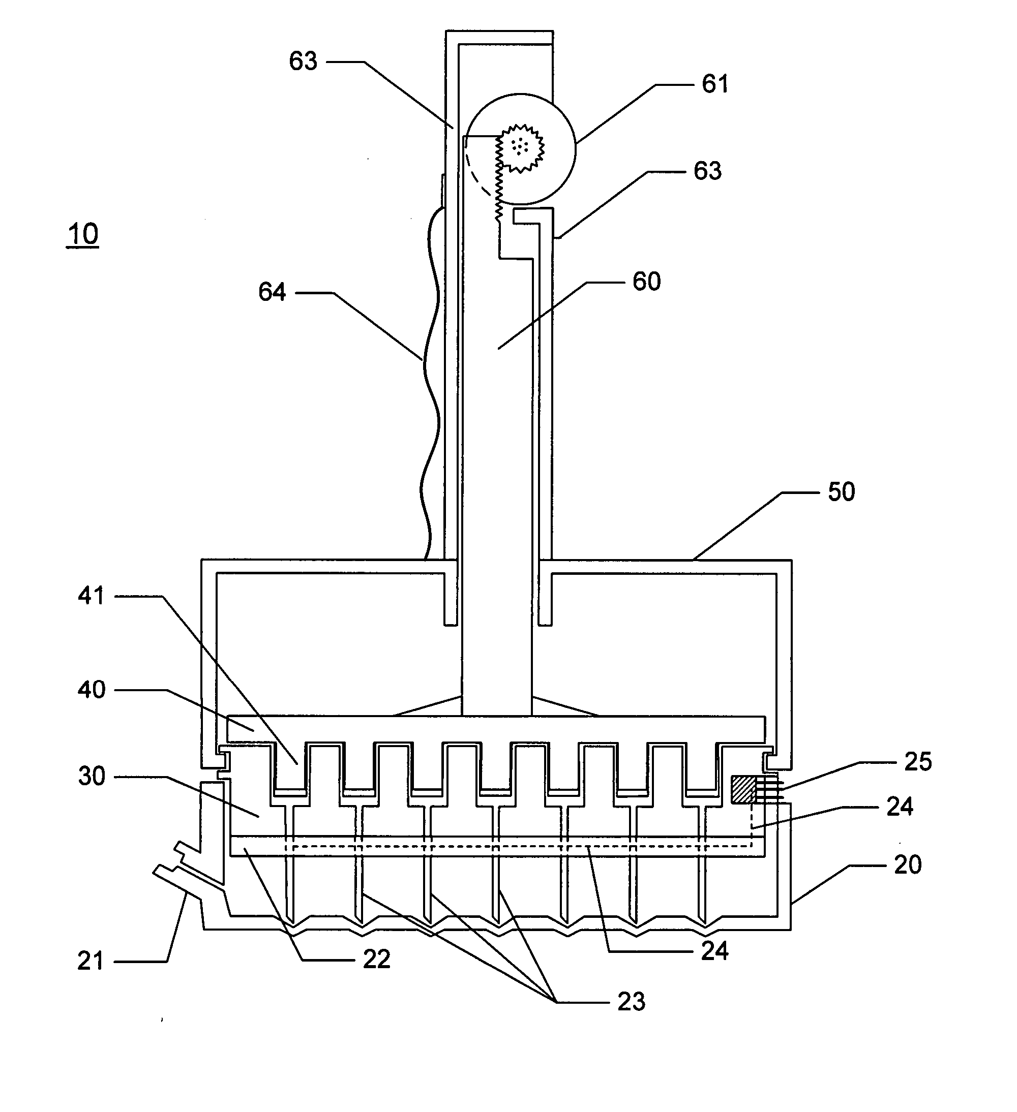
[0061] Turning now to a disclosure of an electroporation device suitable to treat margin beds of resected tumors, in a first embodiment the device is intended to provide the capability to deliver to the margin tissue an anticancer agent, e.g., for example, Bleomycin, evenly distributed throughout the margin bed. In a second embodiment, the device provides for the capability of delivering to the margin tissue a plurality of pulses of electric energy sufficient to cause electroporation of cells throughout the margin bed to a depth of between 1 and 1.5 cm.
[0062] In a third embodiment, the invention device can provide for delivery of the electroporation pulses to a substantial portion of the margin bed, if not all of the margin bed (depending upon the relative dimensions of the device and the tumor), by a single placement of the invention device such that in order to provide electroporating pulses to the entire margin bed the device should preferably only need to be placed once.
[0063]...
PUM
| Property | Measurement | Unit |
|---|---|---|
| Dielectric strength | aaaaa | aaaaa |
| Dielectric strength | aaaaa | aaaaa |
| Dielectric strength | aaaaa | aaaaa |
Abstract
Description
Claims
Application Information