In many cases, the pain severely limits a person's functional ability and
quality of life.
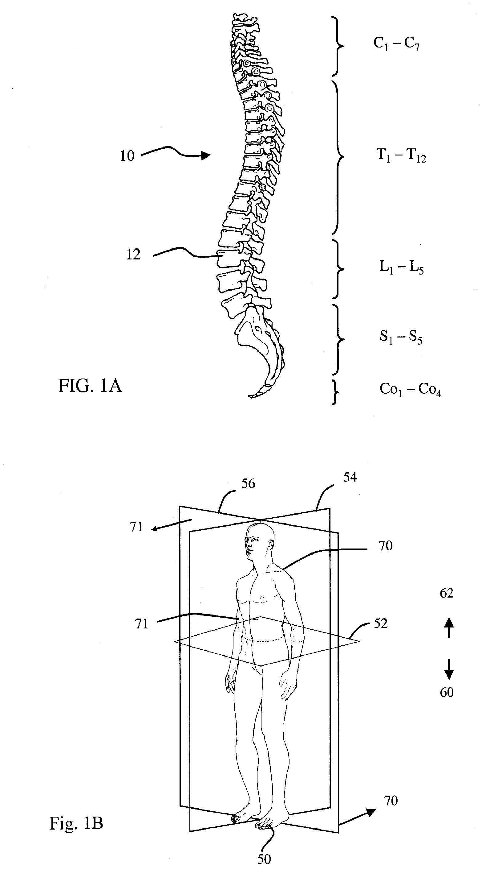
Through
disease or injury, the vertebral bodies, intervertebral discs, laminae,
spinous process,
articular processes, or facets of one or more spinal vertebrae can become damaged, such that the vertebrae no longer articulate or properly align with each other.
This can result in an undesired
anatomy, loss of mobility, and pain or discomfort.
Additionally, there is a substantial
impact on the productivity of workers as a result of lost work days.
Depending upon the extent of the decompression, the removal of support structures such as the
facet joints and / or connective tissues (either because these tissues are connected to removed structures or are resected to access the
surgical site) may result in
instability of the spine, necessitating some form of supplemental support such as
spinal fusion, discussed above.
However, currently available diagnostic techniques provide measurement data that is imprecise and often inconclusive which results in an inability to detect many types of pathologies or accurately assess pathologies that might be considered borderline.
As a result, a significant number of patients having
joint problems remain undiagnosed and untreated using current techniques, or worse are misdiagnosed and mistreated due to the poor
clinical efficacy of these techniques.
For example, currently available techniques for conducting spinal kinematic studies are often unable to determine whether a joint dysfunction is a result of the internal joint structure per se, or whether the dysfunction is a result of, or significantly impacted by, the surrounding muscular tissue.
Additionally, there are no reliable techniques for identifying
soft tissue injury.
Additionally, the level of entrenchment of
muscle guarding behavior cannot currently be determined.
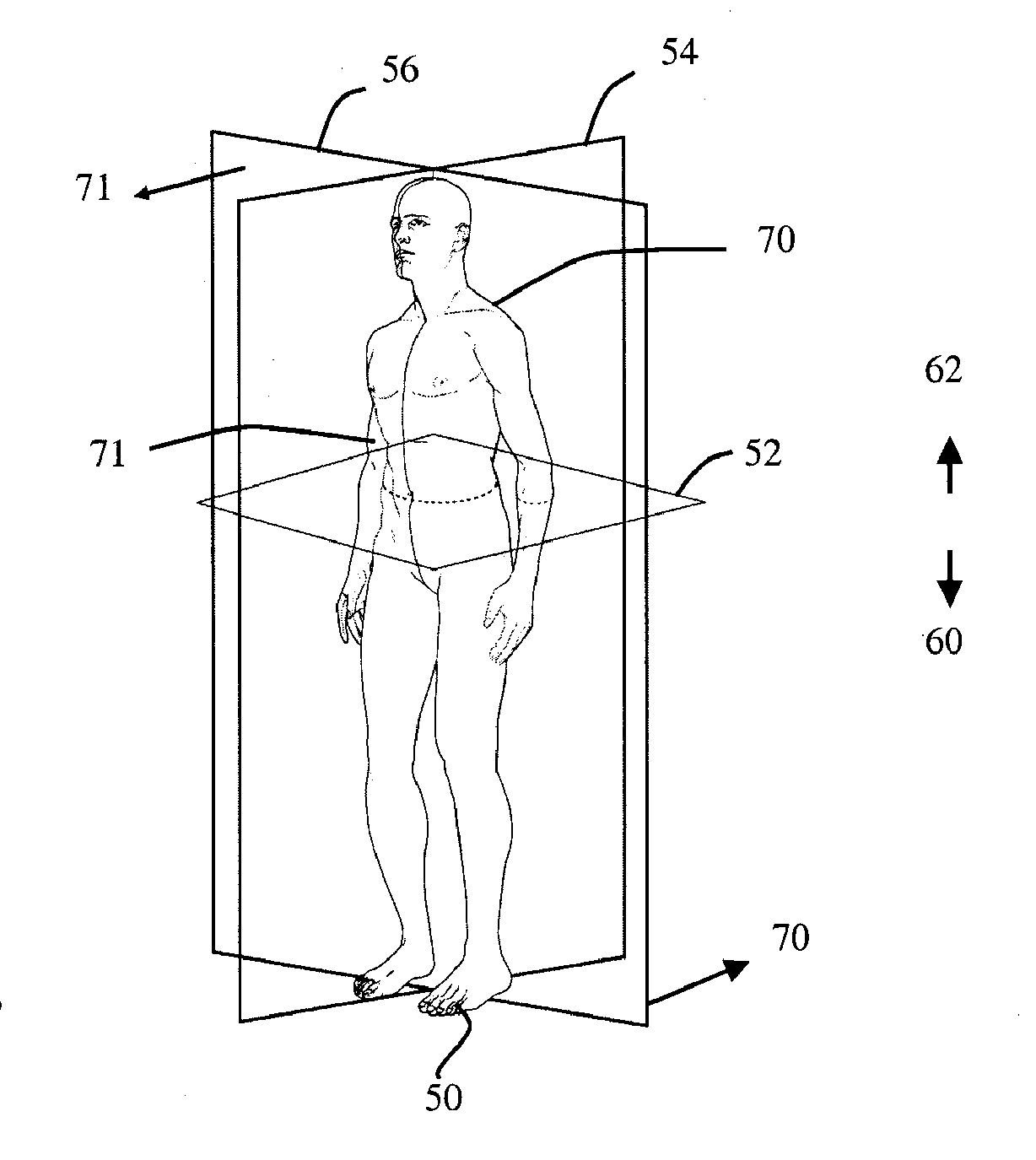
Certain painful movements occur during
joint rotation when the plane of rotation is somewhere between these two postures.
There has not however, been a corresponding improvement in the
diagnostic methods used to identify proper candidates for these interventions.
As a result, the
potential impact and utility of the improvements in orthopedic intervention has been limited.
However, these applications yield poor diagnostic performance with an unacceptably high proportion of testing events yielding either inconclusive or false positive / false negative diagnostic results (Lawrence, J. S. (1969) Annals of Rheumatic Diseases 28: 121-37; Waddell, G.
These references do not, however, provide for any form of measurement or identification of objectively defined motion abnormalities, and therefore is of very limited diagnostic value other than in the detection of grossly and visibly obvious abnormalities that would be detectable using
static image analysis methods.
Without any quantitative or
objective measurement parameters defined, it is impossible to utilize such approaches in comparative analyses across wide populations of subjects, which is required for the purpose of the producing definitive diagnostic interpretations of the results as being either “normal” or “unhealthy”.
Further, there have been no diagnostically useful validations of qualitative motion patterns that are generally absent in non-sufferers but present in subjects suffering from known and specific joint functional derangements or symptoms, or vice versa.
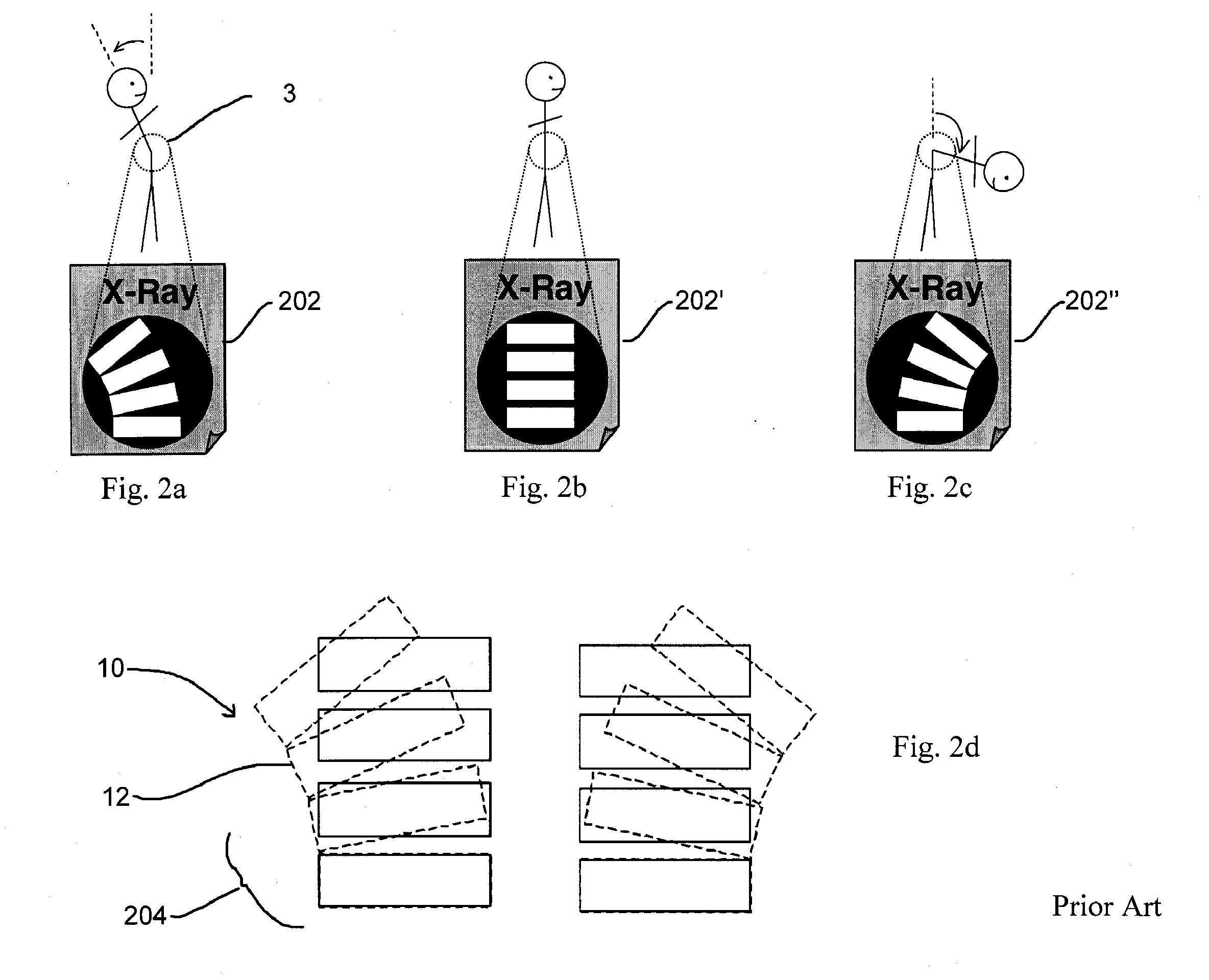
A method for determining
vertebral body positions using
skin markers was developed (Bryant (1989) Spine 14(3): 258-65), but could only measure joint motion at
skin positions and could not measure the motion of structures within the joint.
This method is, however, inherently impractical for clinical diagnostic use.
However RSA requires the surgical implantation of these markers into subjects' internal joint structures, requires the use of two radiographic units simultaneously, and requires a highly complicated calibration process for every
single test, and therefore is too invasive and too cumbersome a process for practicable clinical application.
Using uncontrolled, weight-bearing motion to derive quantitative measurements of joint motion confounds the diagnostic interpretation of such measurements so as to render them diagnostically useless.
Not controlling the motion that is being studied introduces variability into these comparative analyses due to differences that exist across testing subjects with respect to each subject's individual
range of motion, symmetry of motion, and regularity of motion.
Taken together, all of these sources of variability serve to confound diagnostic conclusions based on comparative analyses by making the ranges of “normal” and those of “abnormal” difficult to distinguish from one another other in a statistically significant way.
Such an inability to distinguish between “normal” and “unhealthy” subjects based on a specific diagnostic measurement renders such a measurement diagnostically useless, as has been the case heretofore in the prior art which has focused on measurements of uncontrolled joint motion measured in subjects in weight-bearing postures and moving their joints through the power of their own muscles and in an uncontrolled fashion.
These electromyographical readings of “unhealthy” subjects were then compared to those of a “normal”
population so as to be able to identify those subjects with abnormal readings, however does not provide for a method to report the results as a degree of departure from an ideal reading, instead can only say whether the reading is “abnormal”.
These methods also do not enable determining which type of
muscle group, motive muscles or weight-bearing muscles, or which combination of
muscle groups could be responsible for any observed abnormal electromyographic measurements.
(Lariviere, C 2000) However, visual landmarking methods to not yield as “standardized” a motion as can be achieved with motion that is mechanically controlled, and measurements of the motion of internal joint structures based on surface motion measurements are too variable to be of significant clinical utility.
This method however does not take into consideration the motion of internal joint structures such that a determination as to the specific cause of joint dysfunction cannot be evaluated.
However this approach studied joint motion that was uncontrolled and required an invasive surgical procedure to place the
metal markers, and thus were neither useful nor feasible for clinical diagnostic application.
This approach however does not address the issue of which discrete
muscle group or groups might account for the difference between activation patterns in subjects with joint dysfunctions and those without.
Furthermore, this method does not take into consideration the internal structural joint motions and thus provides an incomplete set of information upon which to draw diagnostic conclusions.
None of the approaches contemplated in the prior art has provided useful, valid, conclusive, and relevant diagnostic results as to the potential presence of joint motion and muscle dysfunctions in a way that controls, standardizes, and measures the tested motion and is clinically practicable and thus potentially able to be integrated into the
standard treatment practice for addressing
joint problems and performance issues.

