Cardiac Motion Artifact Suppression Using ECG Ordering

a motion artifact and ordering technology, applied in the field of magnetic resonance imaging, can solve the problems of increasing scan time, limiting the clinical usefulness of this technique, and not making maximum use of contrast bolus, and achieve the effect of increasing scan tim

Active Publication Date: 2007-12-13
CORNELL RES FOUNDATION INC
View PDF56 Cites 6 Cited by
  • Summary
  • Abstract
  • Description
  • Claims
  • Application Information

AI Technical Summary

Benefits of technology

[0007]The techniques described herein provide a robust ECG ordering for contrast enhanced MRA or MRI without increasing scan time. The ECG signal (representing the cardiac cycle) is monitored in real time. The central k-space views are acquired only in the mid-diastolic rest period of the cardiac cycle. Peripheral views are acquired at any other time, allowing continuous data acquisition. This ECG ordering technique allows a flexible acquisition matrix and is robust against ECG signal imperfections.

Problems solved by technology

However, in the thorax, image quality may suffer from respiratory and cardiac motion and vascular pulsation, which have the potential to cause major ghosting and blurring artifacts, thereby limiting the clinical usefulness of this technique.
However, because these techniques use only a portion of the cardiac cycle, they increase scan time and do not make maximum use of the contrast bolus.

Method used


Image

  • Cardiac Motion Artifact Suppression Using ECG Ordering
  • Cardiac Motion Artifact Suppression Using ECG Ordering
  • Cardiac Motion Artifact Suppression Using ECG Ordering

Examples


Embodiment Construction

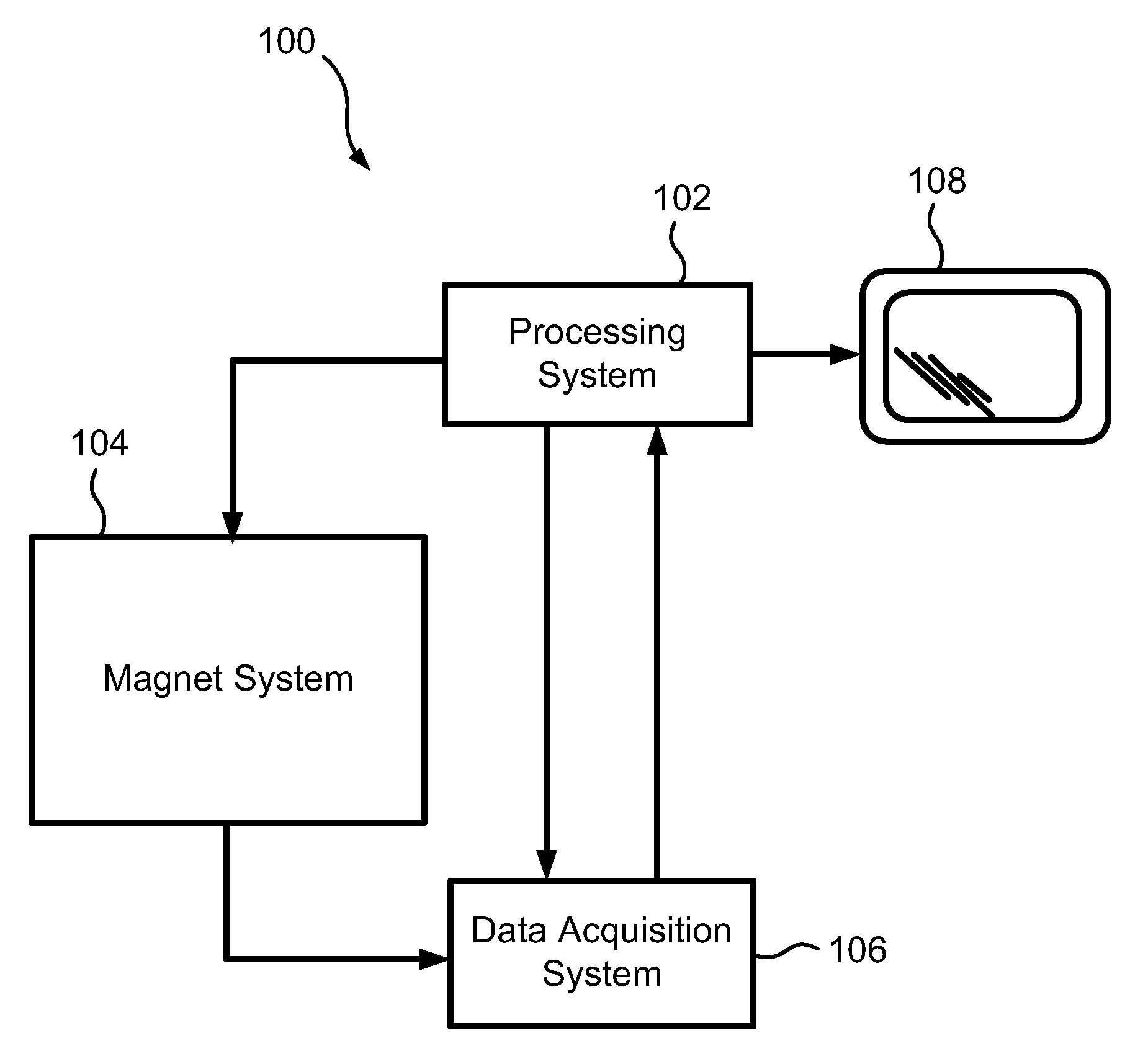
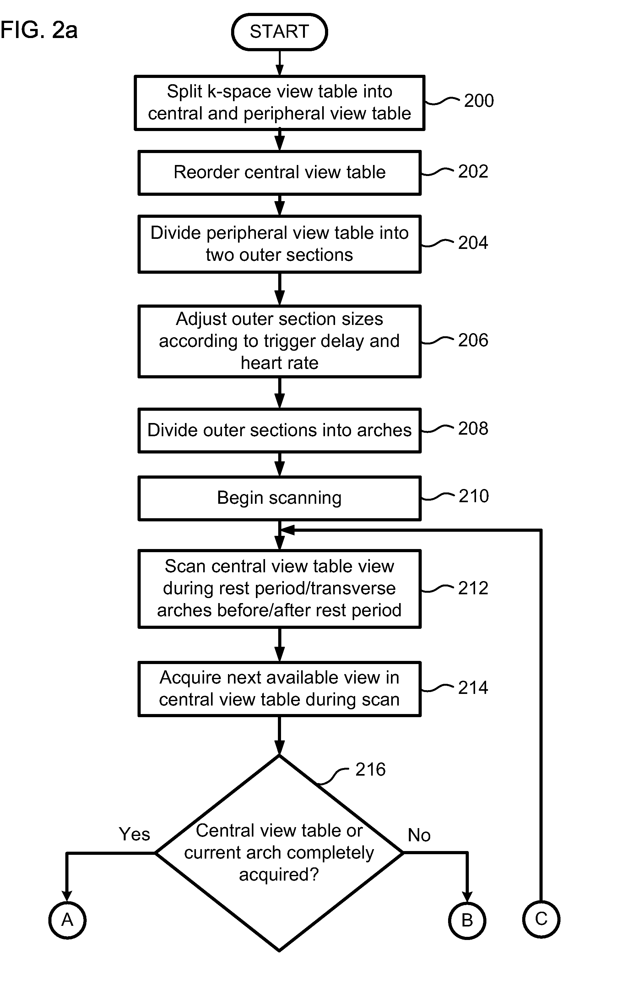
[0031]Described herein is an ECG ordering technique for contrast enhanced magnetic resonance angiography (MRA) and / or imaging (MRI) that combines the advantages of fast continuous scanning, recessed elliptical-centric view ordering and cardiac phase specific acquisition of the central part of k-space. While the ECG ordering technique can be used with MRA and MRI, contrast enhanced MRA shall be used to describe the ECG ordering technique with the understanding that the technique may be used with MRI. The technique suppresses major ghosting and blurring artifacts caused by vascular pulsation and cardiac motion and does so without limiting the acquisition matrix or prolonging scan time. The ECG ordering technique allows a higher resolution MRA to be acquired within the same time frame compared to conventional techniques.

[0032]The data described herein below shows that ECG ordered contrast enhanced magnetic resonance angiography is successful in suppressing ghosting artifacts caused by ...


PUM


Abstract

Described is a robust electrocardiogram (ECG) ordering technique of k-space for breath hold contrast enhanced magnetic resonance angiography (CE-MRA) that acquires the central part of k-space in a motion-free portion of diastole and fills in from the periphery of k-space at all other times. To make maximal use of the contrast enhancement, data is acquired continuously even when the ECG signal is lost. The ECG signal is monitored in real time. The ECG ordering technique allows a flexible acquisition matrix and is robust against ECG signal imperfections. The ECG ordering technique allows thoracic and pulmonary magnetic resonance angiography with a higher resolution when compared to the conventional gated sequence.

Description

CROSS-REFERENCE TO RELATED PATENT APPLICATIONS[0001]This patent application claims the benefit of U.S. Provisional Patent Application No. 60 / 745,298, filed Apr. 21, 2006, the entire disclosure of which is incorporated by reference in their entirety herein.STATEMENT REGARDING FEDERALLY SPONSORED RESEARCH AND DEVELOPMENT[0002]This invention was made in part with Government support under Grant Number HL060879 awarded by the National Institute of Health. The Government may have certain rights in this invention.FIELD OF THE INVENTION[0003]This invention generally relates to magnetic resonance imaging.BACKGROUND OF THE INVENTION[0004]Magnetic resonance imaging (MRI) is the method of choice for noninvasive diagnosis of soft tissue disease in humans, and has wide applications in cardiovascular diseases. For example, contrast enhanced magnetic resonance angiography (CE-MRA) allows a high resolution three dimensional depiction of the vasculature. However, in the thorax, image quality may suff...

Claims


Application Information

Patent Timeline
Patent Type & Authority Applications(United States)
IPC IPC(8): A61B5/055
CPCA61B5/0263A61B5/0402A61B5/055A61B5/7285G01R33/5673G01R33/5601G01R33/5635G01R33/56509G01R33/4818A61B5/318
Inventor SPINCEMAILLE, PASCALWANG, YIPRINCE, MARTIN R.
Owner CORNELL RES FOUNDATION INC
Why Eureka
Try Eureka