
Method for detecting a methylation pattern

a methylation pattern and detection method technology, applied in the field of methylation pattern detection, can solve the problems of methylation information loss in amplification reaction, many conventional detection methods based on hybridization techniques, and difficult detection,

Inactive Publication Date: 2008-03-13
EPIGENOMICS AG
View PDF1 Cites 4 Cited by
  • Summary
  • Abstract
  • Description
  • Claims
  • Application Information

AI Technical Summary

Benefits of technology

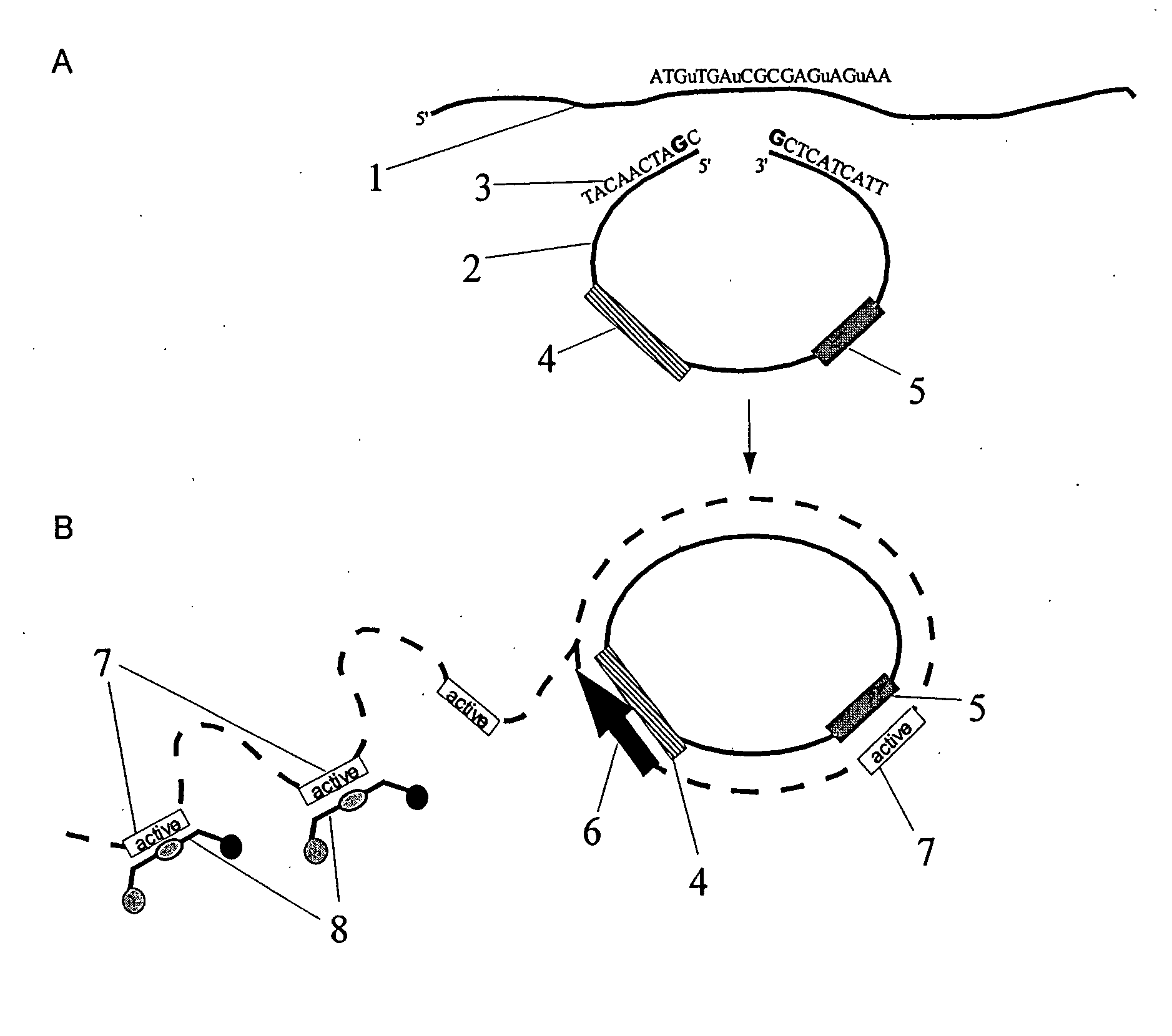
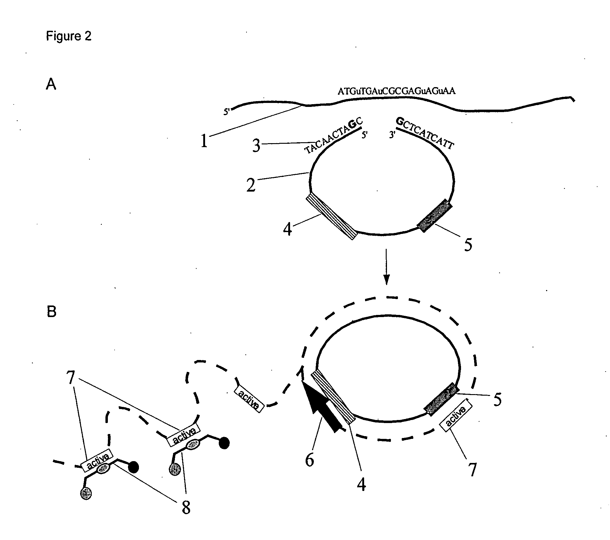
The invention is a method to detect the presence of methylation patterns in nucleic acids. The method involves converting the nucleic acid in a way that allows for the detection of methylation patterns. This can be done by either chemically or enzymatically treating the nucleic acid to prevent changes in unmethylated cytosine. The treated nucleic acid is then brought into contact with a zymogen, which generates a nucleic acid molecule with catalytic activity. The presence or absence of methylation patterns can be detected and quantified. The method can be used with various sources of nucleic acid, such as tissue samples or body fluids. The conversion can be carried out using different methods, such as chemical or enzymatic treatment, and the use of a zymogen. The method allows for the detection and quantification of methylation patterns in nucleic acids.

Problems solved by technology

The detection of methylation is, however, difficult because cytosine and 5-methylcytosine exhibit identical base-pairing behavior.
Many of the conventional detection methods, based on hybridization techniques, do not have the capacity to distinguish between cytosine and 5-methylcytosine.
Furthermore, the methylation information during an amplification reaction, using the polymerase chain reaction (PCR), is completely lost.
However, there exists a problem: that is, in addition to the DNA with methylation patterns typical of a disease-state, one finds a large amount of DNA of the same sequence but with a different methylation pattern.
The applicability of these real-time methods for methylation analyses is, however, limited.
However, in contrast to conventional real-time-PCR methods that which are used for mutational analyses, this method possesses no advantages.
The transfer of the known mutational analysis method for use in methylation analyses cannot be easily performed, since bisulfite-treated DNA is different chemically and physically from genomic DNA in multiple ways; this includes, for example: the length, the complexity, base composition, and the structure: As a result of the bisulfite conversion, the DNA becomes highly fragmented.
To achieve complete conversion, however, correspondingly long reaction times are necessary.
Therefore, the design of specific primers or probes for bisulfite-treated DNA is far more difficult than for genomic DNA.
Methods to analyze genomic DNA are, therefore, off hand not adaptable for the analysis of bisulfite-treated DNA.
An exact quantification of a mutation is often unnecessary because the mutation appears in the DNA of all cells being sampled.
Moreover, the analysis of cytosine methylation particularly from body fluids poses the following problem: besides the DNA with a specific methylation pattern from tumors being located in the body fluids, the body fluids also contain an abundance of DNA that originates from natural breakdown and ageing processes.

Method used


Image

  • Method for detecting a methylation pattern
  • Method for detecting a methylation pattern
  • Method for detecting a methylation pattern

Examples


example 2

Quantification of Methylation of Exon1 in the GSTp1 Gene Using Real-Time PCR

[0203] The previously described PCR reaction in Example 1 is not carried out in a real time duplex reaction, but in individual real-time PCR reactions for methylated- and aggregate-DNA.

example 3

Quantification of Methylation of Exon1 in the GSTp1 Gene Using PCR

[0204] The previously described PCR reaction in Examples 1 and 2 are carried out in a conventional PCR apparatus. The analysis is carried out using endpoint determination of the fluorescent signal as elucidated in WO 2005 / 098035.


PUM

PropertyMeasurementUnit
temperatureaaaaaaaaaa

Abstract

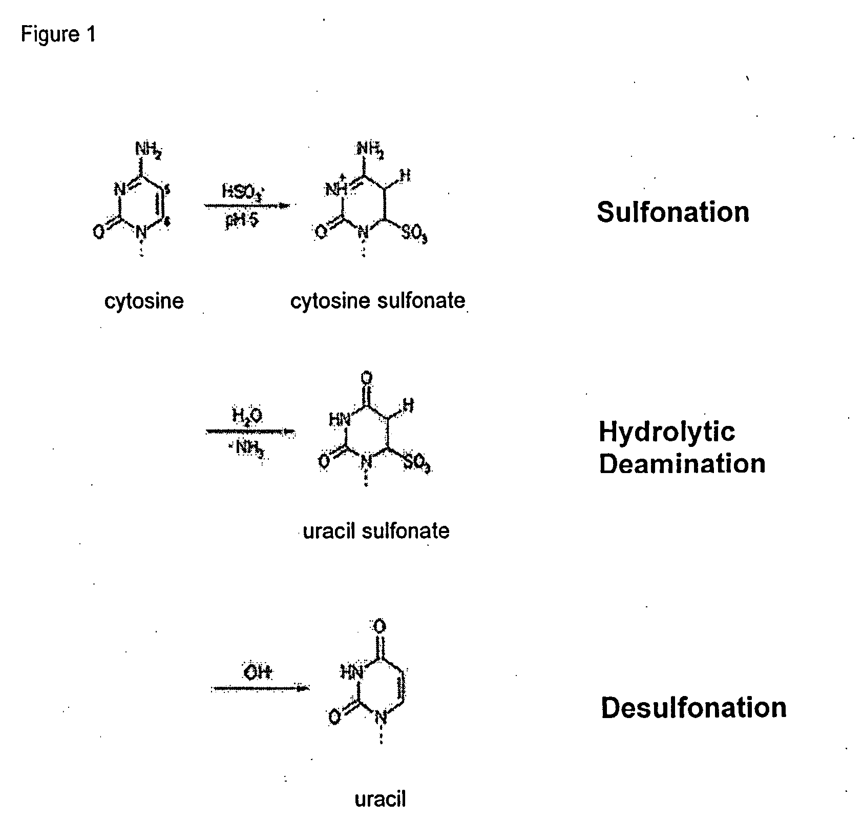
The present invention relates to a method for detecting the presence of one or more methylation patterns. It comprises the conversion of nucleic acids and a catalytic nucleic acid activity. Here, the conversion of the nucleic acid is characterized such that the 5-methylcytosine remains unchanged while the unmethylated cytosine is converted into uracil or another base, which can be distinguished from cytosine in its base-pairing behavior.

Description

[0001] This application incorporates by reference German Appln. No. 10 2006 035 600.4, for which foreign priority is claimed in this application. [0002] The present invention relates to a process for detecting a methylation pattern in a nucleic acid. [0003] In this application, various publications are being cited. The disclosure of the publications is herewith included in the description to describe the relevant state of the art more fully in this application. BACKGROUND OF THE INVENTION [0004] 5-methylcytosine is the most covalently modified base in the DNA of eukaryotic cells. It plays an important biological role, for example, in transcription regulation, genetic imprinting, and in tumorigenesis (for an overview: Millar et al.: Five not four: History and significance of the fifth base. In: The Epigenome, S. Beck and A. Olek (eds.), Wiley-VCH Verlag Weinheim 2003, P. 3-20). The identification of 5-methylcytosine as an integral part of genetic information is, therefore, of great i...

Claims


Application Information

Patent Timeline
Patent Type & Authority Applications(United States)
IPC IPC(8): C12Q1/68C07H21/00G01N33/50
CPCC12Q1/6827Y10T436/143333C12Q2531/125C12Q2523/125C12Q2521/337
Inventor BERLIN, KURTDISTLER, JUERGENTETZNER, REIMO
Owner EPIGENOMICS AG
Learn More