Ultrasonic imaging apparatus, a method for displaying a diagnostic image, and a medical apparatus

a diagnostic image and ultrasonic imaging technology, applied in ultrasonic/sonic/infrasonic diagnostics, instruments, tomography, etc., can solve the problems of difficult for an operator to find abnormalities based on difficult for an operator to find changes in the measured waveform, etc., to achieve accurate diagnosis

Active Publication Date: 2008-03-27
TOSHIBA MEDICAL SYST CORP
View PDF10 Cites 12 Cited by
  • Summary
  • Abstract
  • Description
  • Claims
  • Application Information

AI Technical Summary

Benefits of technology

[0015]According to the relevant art, the ultrasonic tomographic image generated by ultrasonically scanning the subject is displayed, and furthermore the measured waveform and the standard waveform are additionally superposed and displayed. This makes it possible to objectively recognize the changes in measured waveforms in the target organ of the subject and thus contribute to an accurate diagnosis.

Problems solved by technology

Therefore, it has been difficult for an operator to find changes that appear in the measured waveform.
Accordingly, it has also been difficult for an operator to find abnormalities based on changes in the measured waveform.
Furthermore, in diagnosis performed by stress echo, abnormal motion is ascertained by reference to an image, so it relies on the experience of an operator, which has made measurement complicated and also makes the shortening of measurement time difficult.

Method used


Image

  • Ultrasonic imaging apparatus, a method for displaying a diagnostic image, and a medical apparatus
  • Ultrasonic imaging apparatus, a method for displaying a diagnostic image, and a medical apparatus
  • Ultrasonic imaging apparatus, a method for displaying a diagnostic image, and a medical apparatus

Examples


embodiment 1

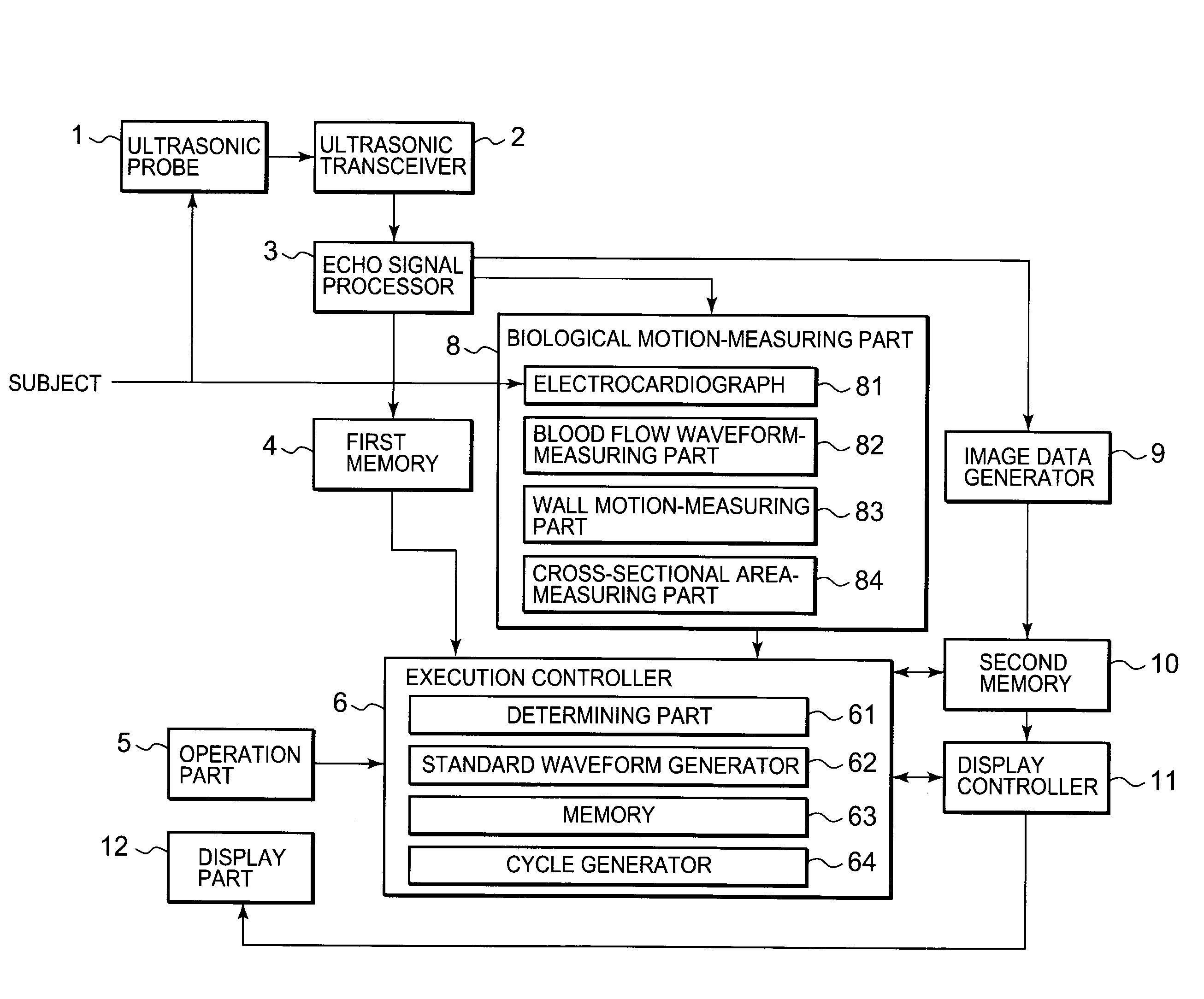
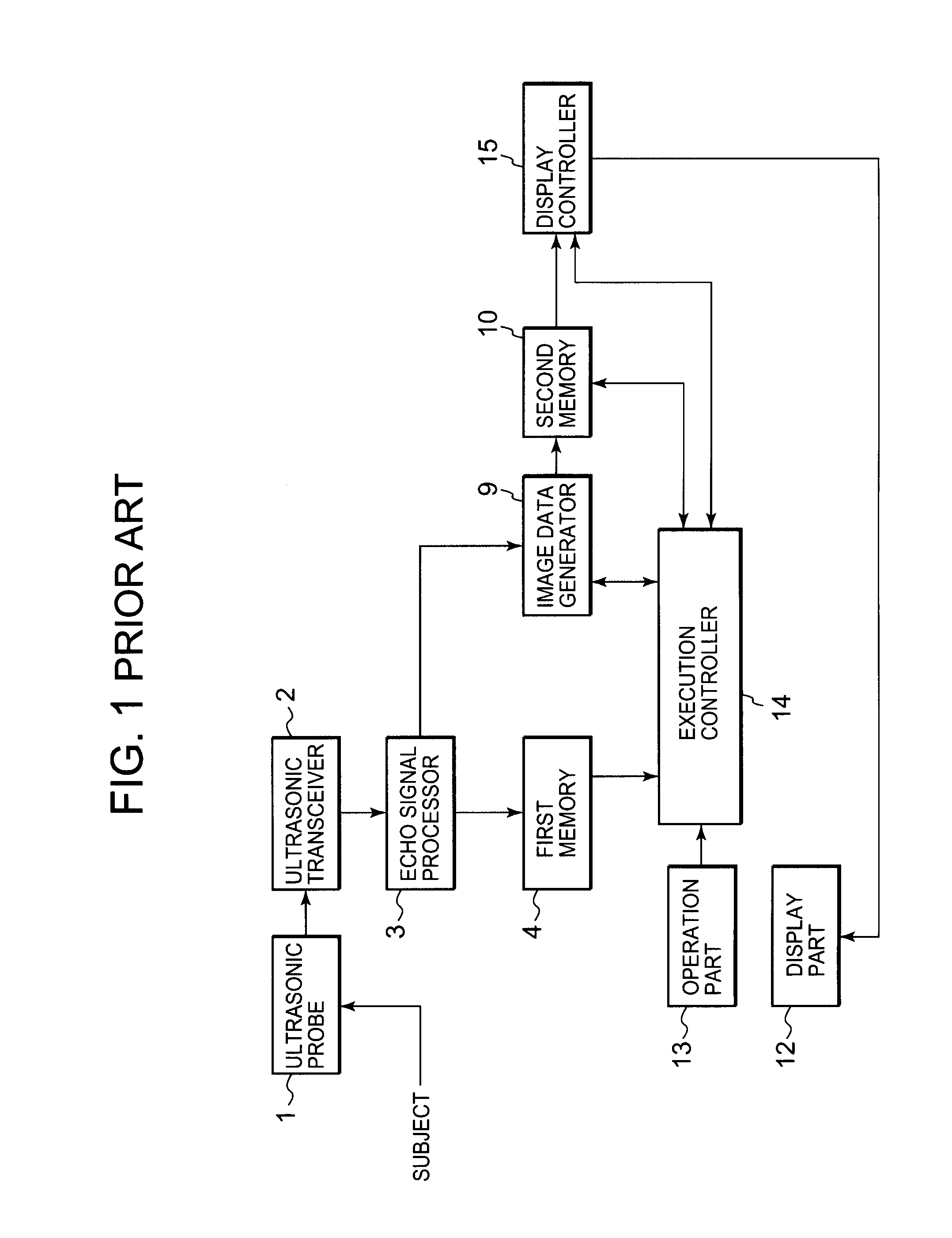
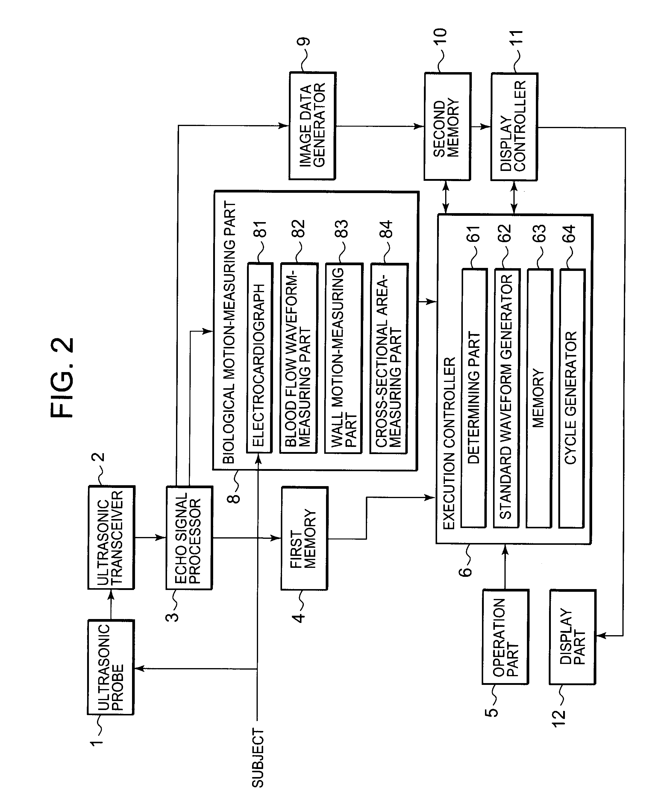
[0028]An ultrasonic diagnostic apparatus according to Embodiment 1 of the present invention will be described below with reference to FIG. 2 to FIG. 8. In FIG. 2, components having the same code as FIG. 1 described in the background have the same function, so the explanation for components having the same code will be omitted. The case in which a biological waveform is used as a measured waveform will be described below. In particular, the case in which an electrocardiographic waveform is used out of the biological waveform will mainly be described.

[0029]FIG. 2 shows a biological motion-measuring part 8 comprising an electrocardiograph 81, a blood flow waveform-measuring part 82 for performing Doppler processing, wall motion-measuring part 83, and a cross-sectional area-measuring part 84. The electrocardiograph 81 generates an electrocardiogram of a subject and obtains an electrocardiographic waveform (FIG. 4 shows a part thereof). The blood flow waveform-measuring part 82 generates...


PUM


Abstract

An ultrasonic transceiver obtains scanning data by transmitting ultrasound to a subject and receiving a reflected wave from the subject. A biological motion part measures, during said scanning, cyclic biological motion of said targeted organ and then calculates a measured waveform that shows motion in relation to time used for said biological motion. An image data generator converts the scanning data into image data shown in a specified coordinate system, and generates an ultrasonic tomographic image. A display controller superposes a standard waveform for a measured waveform, and a measured waveform for the latest cycle, so as to match a specified time phase for each waveform, and causes a display part to display these with the ultrasonic tomographic image.

Description

BACKGROUND OF THE INVENTION[0001]1. Field of the Invention[0002]The present invention relates to an ultrasonic imaging apparatus for generating and displaying an ultrasonic tomographic image by ultrasonically scanning a cross-section of a subject, as well as detecting abnormal motion.[0003]2. Description of the Related Art[0004]An ultrasonic imaging apparatus is capable of obtaining an image in real-time showing, for example, a heartbeat simply by placing an ultrasonic probe on the surface of a subject. Therefore, the ultrasonic imaging apparatus has been used for diagnosing abnormal motion of the target organ, such as abnormality of the diastolic / systolic motion of a heart. The ultrasonic imaging apparatus is safer than other apparatus such as an X-ray diagnostic apparatus, an X-ray CT apparatus, or an MRI apparatus, so it can repeat examinations. Further, the ultrasonic imaging apparatus has a small system scale, so it is simple in that, for example, examinations can be easily per...

Claims


Application Information

Patent Timeline
Patent Type & Authority Applications(United States)
IPC IPC(8): A61B8/13
CPCA61B5/0402A61B8/06A61B8/08G01S7/52087A61B8/13A61B8/465G01S7/52074A61B8/0883A61B5/318
Inventor GUNJI, TAKAYUKI
Owner TOSHIBA MEDICAL SYST CORP
Why Eureka
Try Eureka