
Apparatus and method for diagnosing ischemic heart disease

a technology ischemic heart disease, applied in the field of ischemic heart disease apparatus and the method field, can solve the problems of high health care cost of this procedure, serious hemorrhaging or large hematoma, and only about 65% of the diagnosis rate of electrocardiogram

Inactive Publication Date: 2008-04-03
KAKIHARA RI ICHIRO
View PDF1 Cites 13 Cited by
  • Summary
  • Abstract
  • Description
  • Claims
  • Application Information

AI Technical Summary

Benefits of technology

[0014]According to the present invention, ischemic heart disease can be diagnosed at a probability roughly equal to that of stress echocardiography without placing a burden on the patient based on echocardiographical images obtained while the patient is at rest.

Problems solved by technology

Therefore, although being rare cases, serious hemorrhaging or large hematomas may be caused.
Also, there is another disadvantage of high health care costs for this procedure.
However, the diagnosis rate when an electrocardiogram is employed is only about 65%, and 10% of these cases, when further assessed by coronary angiography, prove to be cases which require surgical operations.
However, this method, which strictly relies on images acquired by echocardiography, requires a considerable amount of experience on the part of each physician, in that it is essential yet technically difficult to keep applying adequate stress to heart and obtain images of constant and stable diagnostic sensitivity.

Method used


Image

  • Apparatus and method for diagnosing ischemic heart disease
  • Apparatus and method for diagnosing ischemic heart disease
  • Apparatus and method for diagnosing ischemic heart disease

Examples


examples

[0059]The present invention was carried out according to the following examples using actual specific cases including normal and stenosed cases.

[0060]The GE Vivid 7 Dimension Version 4.1.0 was used for the ultrasound system, and images were recorded with the patients at rest. Data was analyzed using EchoPAC PC Dimension Version 4.1.0 off-line.

[0061]The images used for analysis were comprised of three cross-sections consisting of a four-chamber view (Ap4ch view), long-axis view (ApLax view) and two-chamber view (Ap2ch view) from an apical approach. The center of the left ventricular anterior wall of the two-chamber view was designated as the left anterior descending coronary artery (LAD) region, the center of the left ventricular inferior wall of the two-chamber view was designated as the right coronary artery (RCA) region, and the center of the left ventricular posterior wall of the long-axis view was designated as the left circumflex coronary artery (LCX) region.

[0062]After designa...


PUM


Abstract

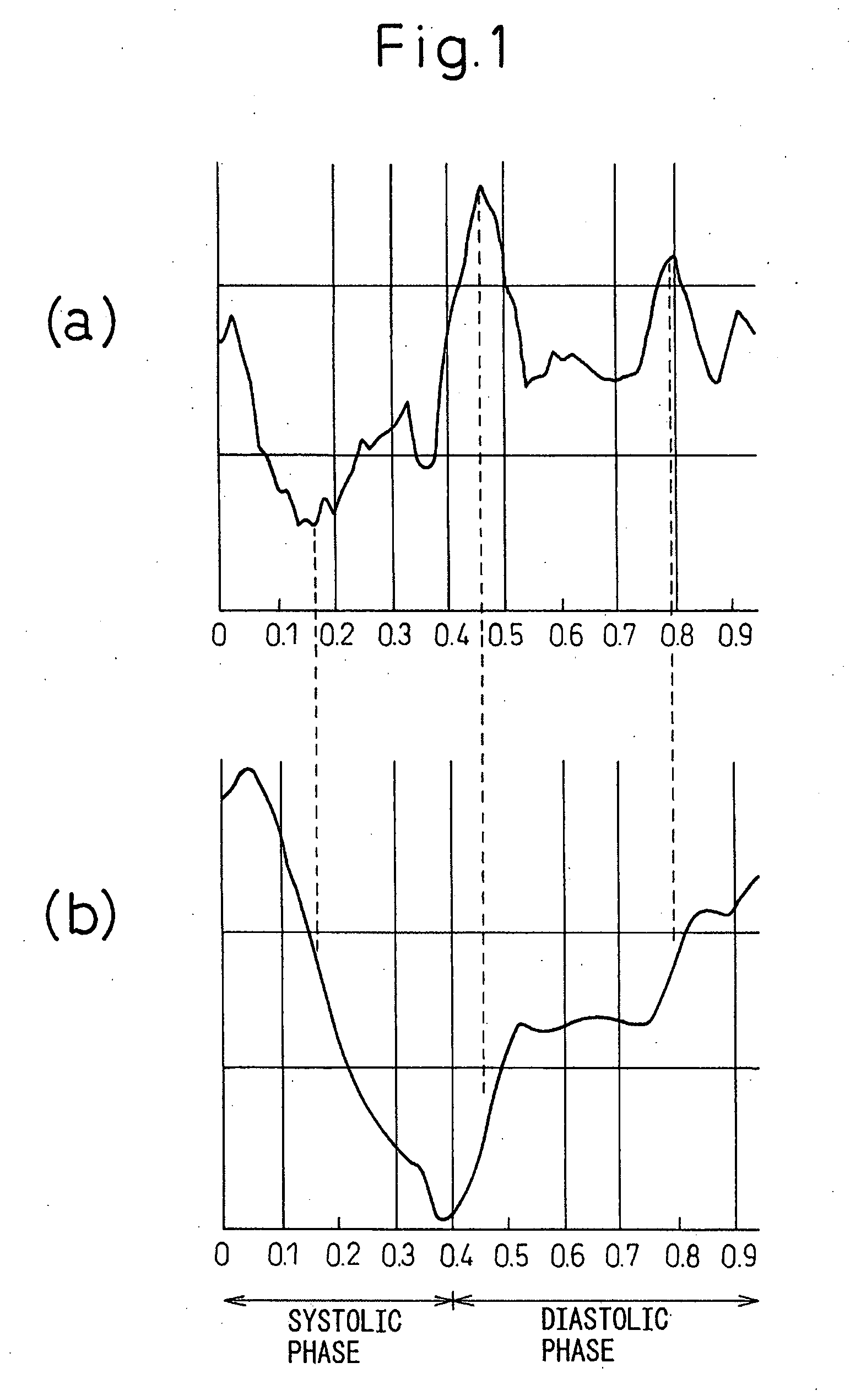
A region of interest is set at a thin layer on the inside of the left ventricular wall for ecocardiographical apical long-axis tomographic images obtained while at rest, and the strain rate of the set region of interest is calculated. The value of a discriminant function is determined on the basis of a plurality of strain rates of an intermediate portion of the systolic phase. Ischemic heart disease is then diagnosed according to the value of the discriminant function.

Description

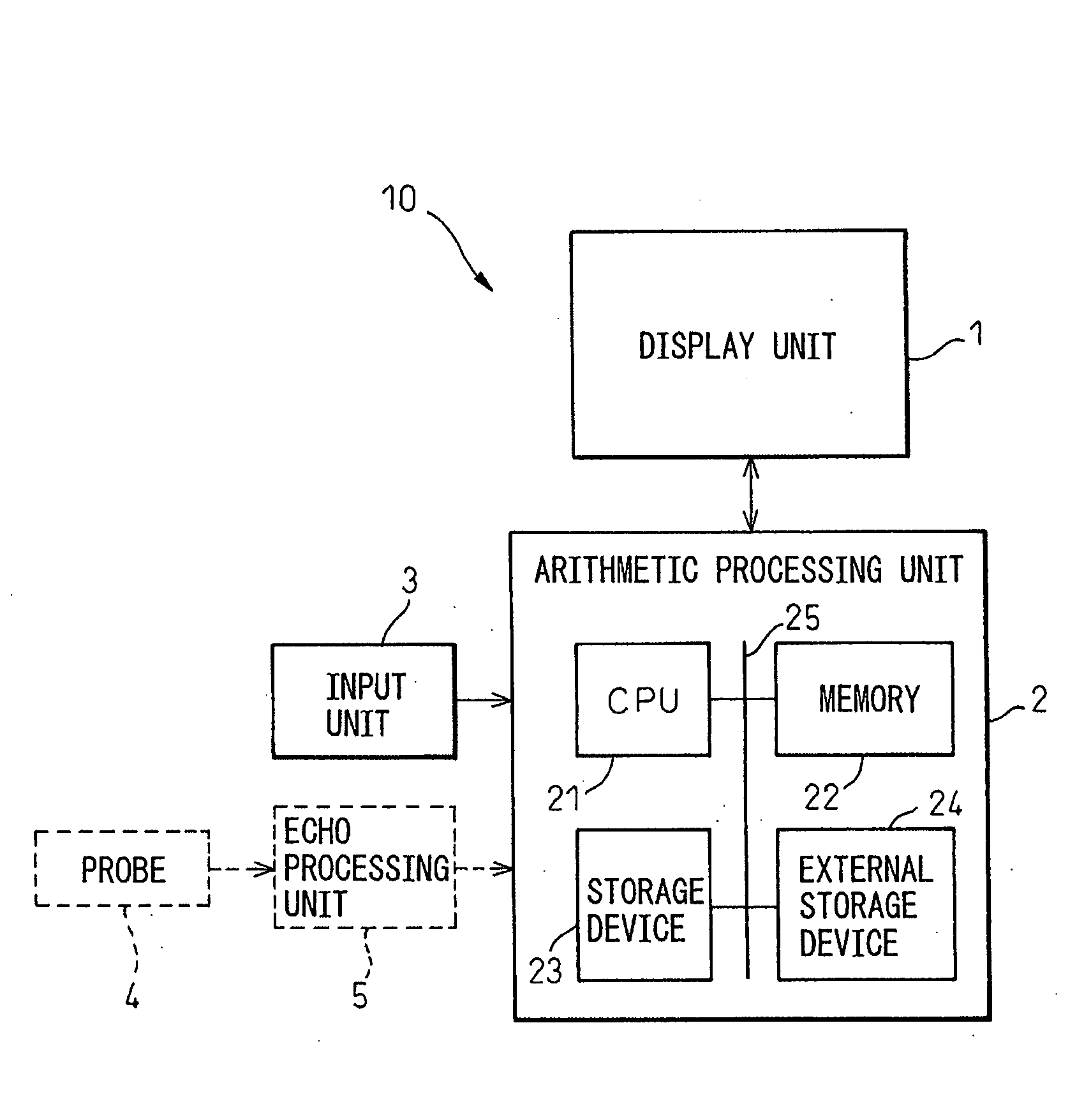
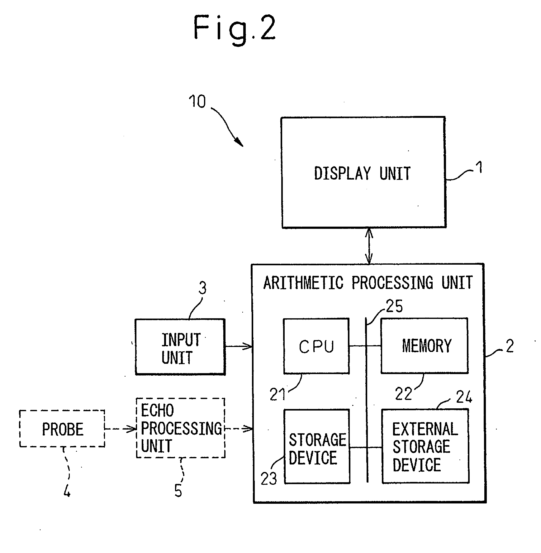
FIELD OF THE INVENTION[0001]The present invention relates to an apparatus and a method that diagnose ischemic heart disease by echocardiography.BACKGROUND OF THE INVENTION[0002]Conclusive diagnosis of ischemic heart disease is given by coronary angiography. The process of coronary angiography consists of insertion of a narrow catheter into a coronary artery from an arm or the groin, injection of a contrast agent for X-ray imagining, and examination of X-rays from multiple directions. The acquired X-ray images are observed macroscopically, and the percentage of stenosis at each portion of the coronary arteries is examined for the assessment of future treatment. This process is wholly based on the operator's experience. Future treatment is then determined, including whether surgical treatment should be performed on the patient.[0003]As stated above, coronary angiography involves the insertion of a foreign body into a blood vessel, and is often performed even on patients with high degr...

Claims


Application Information

Patent Timeline
Patent Type & Authority Applications(United States)
IPC IPC(8): A61B8/14G06F19/00A61B8/08
CPCA61B8/08A61B8/0883A61B8/485G06T2207/30048G06T2207/10072G06T2207/10132G06T7/0012
Inventor KAKIHARA, RI-ICHIRO
Owner KAKIHARA RI ICHIRO
Learn More