Apparatus and Methods for Performing Brain Surgery

a brain surgery and apparatus technology, applied in the field of apparatus and methods for brain surgery, can solve the problems of high invasiveness of brain surgery, difficult diagnosis and treatment of brain conditions, and inability to adapt to all situations,

Inactive Publication Date: 2008-05-08
NICO CORP
View PDF26 Cites 104 Cited by
  • Summary
  • Abstract
  • Description
  • Claims
  • Application Information

AI Technical Summary

Benefits of technology

[0011]Traditional methods are used to incise and retract soft tissue of the scalp covering the skull. A hole is made in the skull, and the dura is opened and retracted to provide access to the brain. In the first method, the stylet or probe is inserted through the obturator longitudinal channel and advanced until a length of the stylet or probe extends out of and beyond the blunt tip of the dilating obturator. The dilating obturator and cannula assembly is held back away from the tissue as the stylet or probe is gently advanced through the brain tissue under both direct vision and positional image guidance until the tip of the stylet or probe is adjacent the target tissue. Once the stylet or probe is placed and the position is confirmed using the image guidance system, the blunt dilating obturator and cannula assembly is slowly and carefully advanced into the brain tissue to atraumatically spread the tissue over the dilating tip and around the cannula while maintaining the position of the stylet or probe as a guide to advancement of the obturator and cannula assembly. A gentle back and forth rotation during insertion may facilitate placement of the obturator cannula assembly. Once the dilating obturator and cannula assembly are correctly positioned adjacent the target tissue, the stylet or probe and dilating obturator are removed, leaving the cannula in place to support and protect the dilated brain tissue. Preferably, the cannula is clear so that the dilated brain tissue may be visually inspected through the walls of the cannula to assure that no damage was caused to surrounding brain tissue during insertion of the device.
[0016]Appropriate surgical instruments are then used to perform surgery upon the target tissue. For example, scissors, graspers and suction tools may be inserted through the cannula, visualizing the tips of the instruments to perform the desired procedure either directly with the naked eye or through a microscope, or indirectly through the endoscope using the endoscope eyepiece or more preferably and camera system to display the image on a monitor. A preferred instrument for debulking brain tissue is a fluidized ultrasonic instrument, such as CUSA (Valleylab, Boulder Colo.) During surgery, monitoring equipment may be used to monitor brain function during surgery to assist the surgeon in understanding the effects of the actions taken during surgery operating on the brain so that the surgery may be terminated in the event an indication of an adverse effect is detected.
[0018]The devices and methods disclosed herein provide numerous advantages in performing brain surgery. Gentle atraumatic dilation of the brain tissue makes it possible to operate further inside the brain than otherwise would be possible utilizing traditional surgical techniques. The disclosed methods and apparatus create an access area to work while simultaneously protecting adjacent brain tissue from inadvertent collateral damage and trauma that might otherwise occur if more traditional surgical techniques were to be utilized. In addition, accessing target tissue through the cannula as contemplated avoids more invasive techniques that involve removing substantial portions of the skull and retracting large portions of the brain to gain access to operate on target tissues. In some cases, the devices and methods may make it possible to operate on target tissue that would, without these devices and methods, otherwise be regarded as inoperable using previously known techniques.

Problems solved by technology

Diagnosis and treatment of conditions affecting the brain are among the most difficult and complex problems that face the medical profession.
Given the difficulty of accessing the brain through the hard bony protective skull the diagnosis and treatment of brain disorders presents unique challenges not encountered elsewhere in the body.
Brain surgery can be highly invasive.
Of course, such techniques are not appropriate for all situations, and not all patients are able to tolerate and recover from such invasive techniques.
It is also known to access certain portions of the brain by forming a hole in the skull, but only limited surgical techniques may be performed through such smaller openings.

Method used


Image

  • Apparatus and Methods for Performing Brain Surgery
  • Apparatus and Methods for Performing Brain Surgery
  • Apparatus and Methods for Performing Brain Surgery

Examples


first embodiment

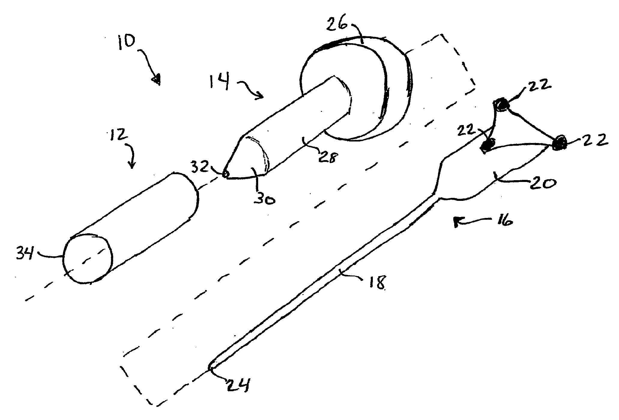
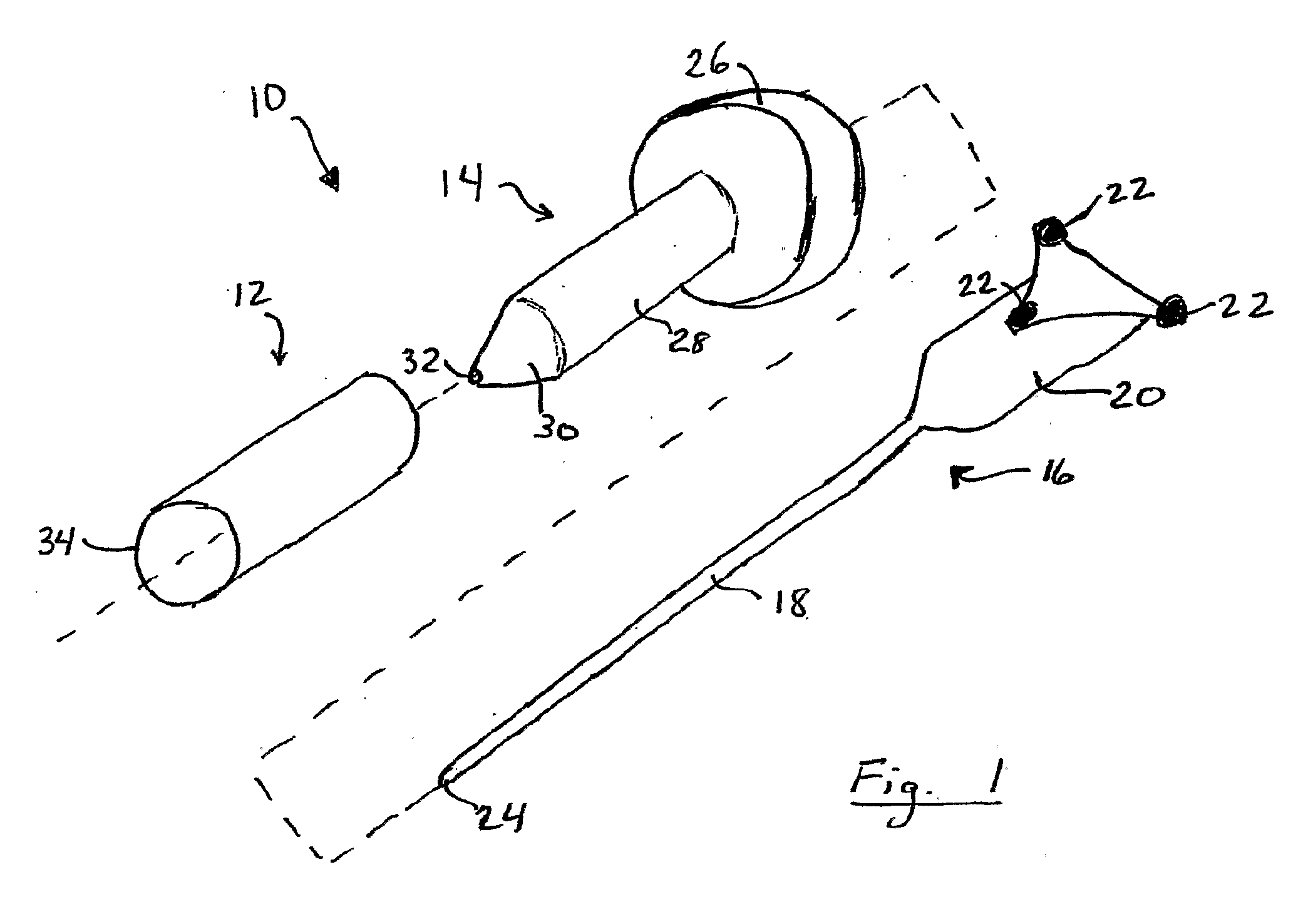
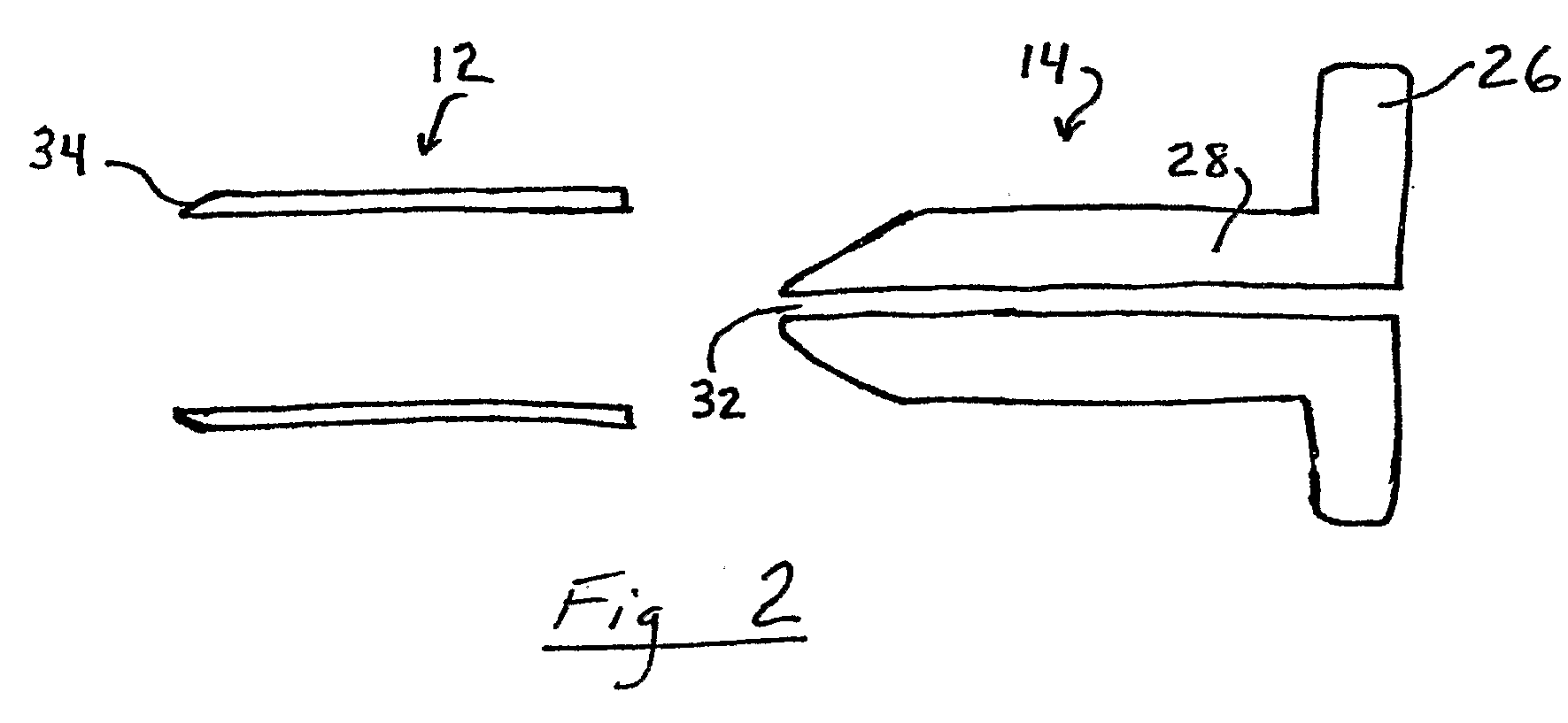
[0032]Referring now to the drawings, FIG. 1 illustrates an apparatus 10 for accessing target tissue within the brain in order to perform brain surgery. The access device includes a cannula 12, a dilating obturator 14 and a stylet or probe 16. Stylet or probe 16 has a small diameter elongated shaft 18, a handle 20 and associated position indicators 22 for an image guidance system. Stylet shaft 18 has a blunt tip 24 that can be inserted into and advanced through brain tissue. In FIG. 1, image guidance position indicators are shown as infrared reflectors of the type use in connection with optical image guidance systems. As shown, the infrared reflectors used with such a system are mounted to the stylet handle in a customary triangular configuration calibrated to identify the tool to the image guidance system. Such imaging systems are available, for example Medtronic Surgical Navigation Technologies (Denver, Colo.), Stryker (Kalamazoo, Mich.), and Radionics (Burlington Mass.).

[0033]Typi...

third embodiment

[0039]FIG. 5 is a perspective view, with parts separated, of an access device 70 for brain surgery. Access device 70 includes cannula 72 with chamfered lead edge 74, and a dilating obturator 76. Dilating obturator 76 includes a handle 78, substantially cylindrical shaft 80 and dilating tip 82, which is preferably conical with a rounded distal tip. Access device 70 does not include apparatus for calibrating the position of the dilating obturator with an image guidance system or a stylet or probe for aiding insertion of the dilating obturator.

[0040]FIGS. 6-10 illustrate the use of the access device 10 of the first embodiment during minimally invasive brain surgery, as will now be described.

[0041]In FIG. 6, a partial cross-section view of the access device 10 with stylet shaft 18 of stylet 16 inserted through an opening 100 formed in a patient s skull 102 through brain tissue until tip 24 of stylet 28 is adjacent target tissue 104. Opening 100 is made in a traditional manner, by incisi...


PUM


Abstract

Less invasive surgical techniques for performing brain surgery are disclosed in which a dilating obturator and cannula assembly is inserted into brain tissue until the obturator tip and cannula are adjacent the target tissue. The obturator is removed and surgery is performed through the cannula. In preferred embodiments the obturator and cannula are placed using image guidance techniques and systems to coordinate placement with pre-operative surgical planning. A stylet with associated image guidance may he inserted prior to insertion of the obturator and cannula assembly to guide insertion of the obturator and cannula assembly. Surgery preferable is performed using an endoscope partially inserted into the cannula with an image of the target tissue projected onto a monitor.

Description

PRIORITY[0001]This application claims the benefit of the priority of co-pending U.S. Provisional application 60 / 623,094, filed Oct. 28, 2004.TECHNICAL FIELD[0002]The present disclosure relates to methods of accessing and performing surgery within the brain.BACKGROUND[0003]Diagnosis and treatment of conditions affecting the brain are among the most difficult and complex problems that face the medical profession. The brain is a delicate soft tissue structure that controls bodily functions through a complex neural network connected to the rest of the body through the spinal cord. The brain and spinal cord are contained within and protected by significant bony structures, e.g., the skull and the spine. Given the difficulty of accessing the brain through the hard bony protective skull the diagnosis and treatment of brain disorders presents unique challenges not encountered elsewhere in the body.[0004]Diagnosis of brain disorders requires clear, accurate imaging of brain tissue through th...

Claims


Application Information

Patent Timeline
Patent Type & Authority Applications(United States)
IPC IPC(8): A61B17/00A61B1/00A61M29/00A61M5/178
CPCA61B1/042A61B2019/5255A61B17/00234A61B17/0293A61B17/3417A61B17/3421A61B19/201A61B19/5212A61B19/5244A61B2017/00907A61B2017/320044A61B2017/3445A61B2019/5206A61B2019/5217A61B1/3132A61B34/20A61B90/11A61B90/361A61B2034/2055A61B2090/306A61B2090/3614
Inventor KASSAM, AMIN
Owner NICO CORP
Why Eureka
Try Eureka