Methods for fat signal suppression in magnetic resonance imaging

a magnetic resonance imaging and fat signal technology, applied in the field of magnetic resonance imaging, can solve the problems of obscuring important pathology, affecting the quality of the acquired image, so as to achieve significant reduction of total acquisition time, high contrast water, and time saving

Active Publication Date: 2008-09-11
CASE WESTERN RESERVE UNIV
View PDF25 Cites 22 Cited by
  • Summary
  • Abstract
  • Description
  • Claims
  • Application Information

AI Technical Summary

Benefits of technology

[0008]More particularly, the systems and methods according to the present invention, in an embodiment, provide for constructing an image from an MRI data set, wherein the MRI data is acquired with a total sampling time (TST) acquisition, with a predetermined timing relationship between echoes during a single acquisition process. Accordingly, the present invention provides significant improvement over traditional 2-point Dixon methods in terms of time savings in data acquisition, while providing desired suppression of fat or water signals. The total acquisition time is significantly reduced as compared to traditional 2-point Dixon methods in comparable sequences. These objectives may be realized using an apparatus to produce MRI images of a scanned objec

Problems solved by technology

In MRI imaging of chemical species such as fat and water within body tissue, the misregistration of fat in acquired magnetic resonance images can seriously degrade the quality of the image and obscure important pathology.
As a diagnostic tool, such off-resonance effects, which may arise from field inhomogeneities, susceptibility or chemical shift, cause artifacts in the acquired images.
Although various fat suppression methods have been conceived, such as using specialized radio-frequency (RF) excitation pulse sequences, such methods have various limitations.
For example, inversion recovery pulse sequences and chemical shift selective excitation p

Method used


Image

  • Methods for fat signal suppression in magnetic resonance imaging
  • Methods for fat signal suppression in magnetic resonance imaging
  • Methods for fat signal suppression in magnetic resonance imaging

Examples


examples

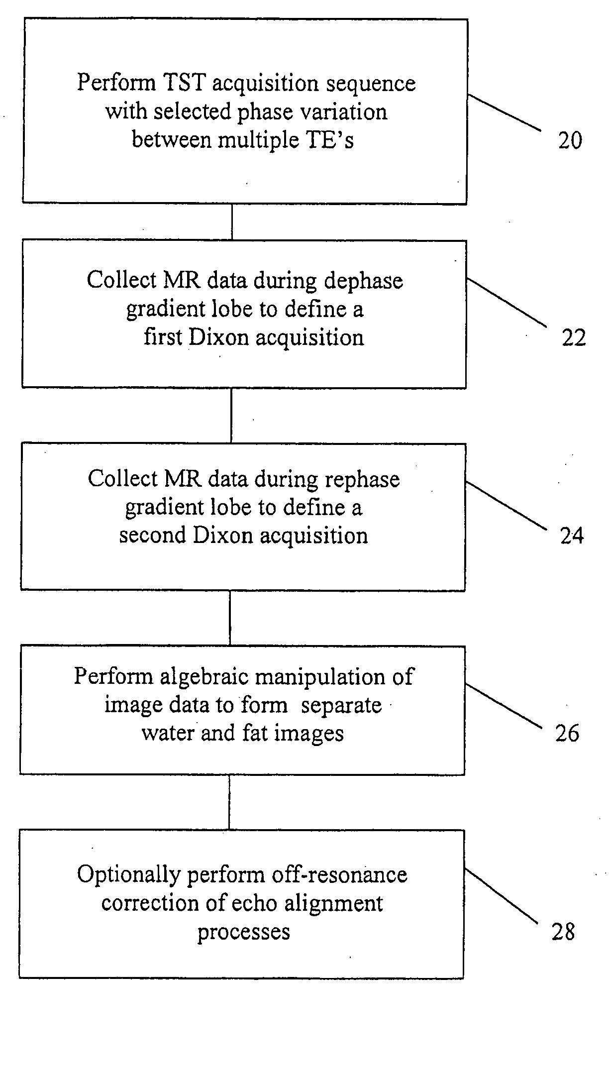
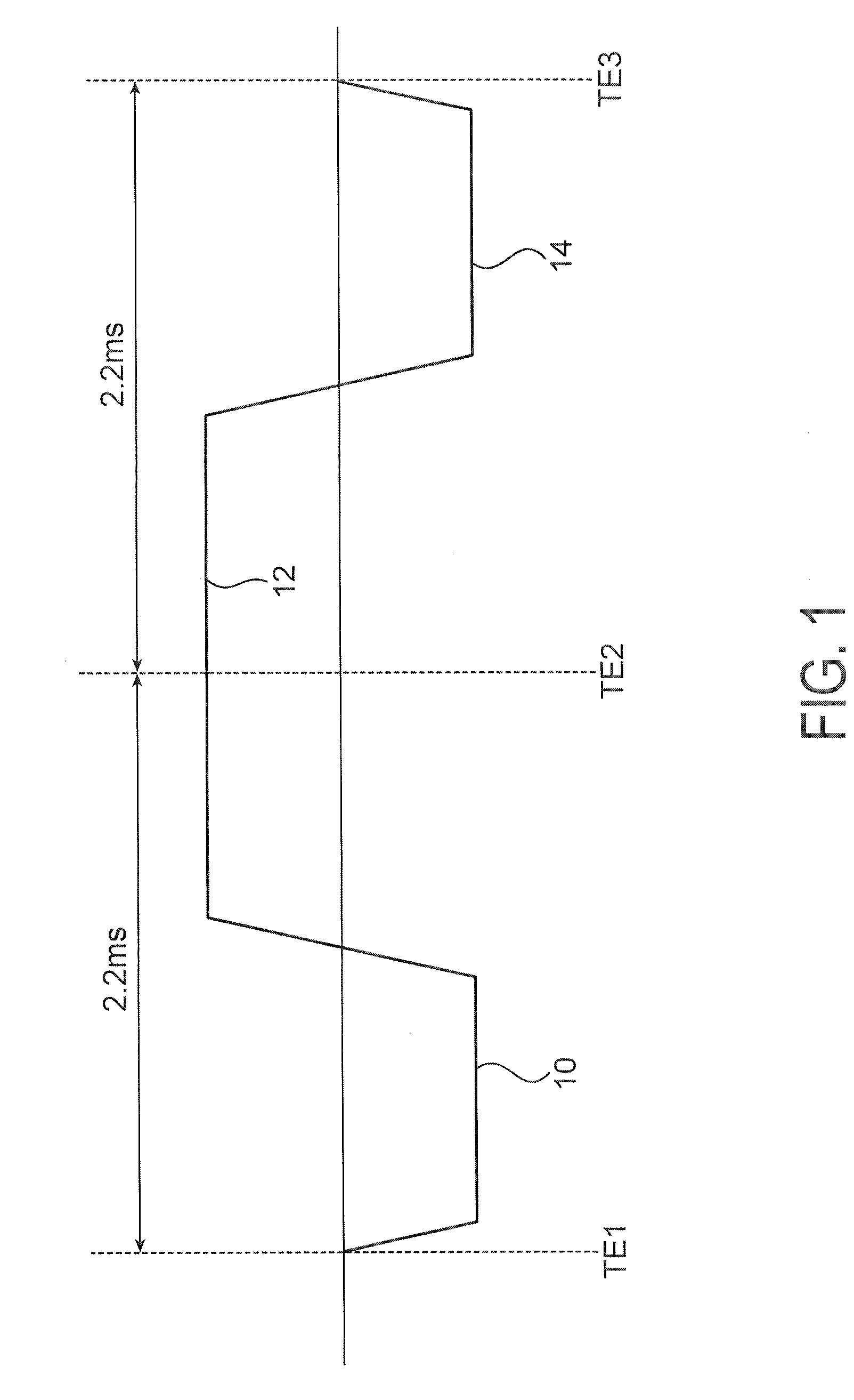
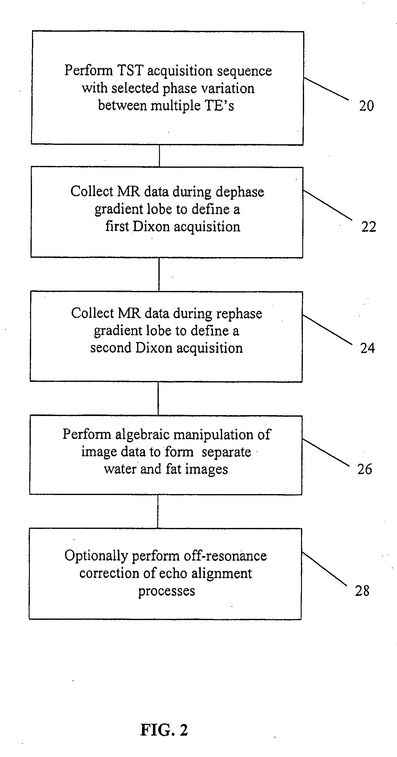
[0026]FIGS. 3A-3C and 4A-4C show the results according to use of the methods of the invention as compared to typical two-point Dixon methods. As a particular example, a FLASH pulse sequence according to FIG. 1 was implemented on a 1.5 T Siemens Sonata MR Scanner, with TE1, TE2 and TE3 selected to allow 180° phase variation in the fat magnetization between each of the three TE's. Imaging parameters used were TE1 / TE2 / TE3=3.4 / 5.6 / 7.8 milliseconds, TR=14 milliseconds, FOV=300 millimeter2, slice thickness=5 millimeters. A second acquisition was collected with TE2=7.8 milliseconds in order to perform traditional, two-point Dixon fat suppression for comparison. Phantom and clinical images were acquired with the TST sequence of FIG. 1, with the dephase / rephase (S1) and readout (S2) k-space data gridded separately using linear interpolation from a measured trajectory. The water and fat images were calculated by adding or subtracting S1 and S2 and then performing 2D Fast Fourier Transform (2D...


PUM


Abstract

The present invention is directed to methods for chemical species signal suppression in magnetic resonance imaging procedures, wherein Dixon techniques are enhanced by continuously sampling techniques. In the invention, k-space data is acquired during the entire period of read gradient associated with a gradient echo pulse acquisition scheme. The invention utilizes a total sampling time (TST) acquisition during the entire read gradient, using three echoes of a TST data set to achieve chemical species separation in both homogenous fields as well as areas of field inhomogeneity. As an example, a continuously sampled rectilinearly FLASH pulse sequence is modified such that the time between echoes was configured to be 2.2 milliseconds, with TE selected to allow 180° phase variation in the fat magnetization between each of the three TE's (TE1, TE2, and TE3). Data collected during the dephase and rephase gradient lobes are defined as a first Dixon acquisition, with data collected by the read gradient lobe being defined as a second Dixon acquisition. Two point Dixon reconstruction techniques are used to form images for each chemical species, such as for generating water and fat images of the scanned object region. Other corrections, such as off-resonance correction may be applied on the image data.

Description

CROSS-REFERENCE TO RELATED APPLICATIONS / INCORPORATION BY REFERENCE[0001]This U.S. Patent Application claims priority to and the benefit of U.S. Provisional Patent Application Ser. No. 60 / 894,070 filed on Mar. 9, 2007 which is incorporated herein by reference in its entirety.FIELD OF THE INVENTION[0002]The present invention relates to a method of fat signal suppression or separation of fat and water signals in MRI imaging of a scanned object region using rectilinear Dixon techniques.BACKGROUND OF THE INVENTION[0003]In MRI imaging of chemical species such as fat and water within body tissue, the misregistration of fat in acquired magnetic resonance images can seriously degrade the quality of the image and obscure important pathology. As a diagnostic tool, such off-resonance effects, which may arise from field inhomogeneities, susceptibility or chemical shift, cause artifacts in the acquired images. The artifacts appear as positional shifts along the read direction in rectilinearly sam...

Claims


Application Information

Patent Timeline
IPC IPC(8): G01R33/483
CPCG01R33/4828G01R33/5614G01R33/5613G01R33/5607
Inventor BOOKWALTER, CANDICE A.SUNSHINE, JEFFREY L.DUERK, JEFFREY L.
Owner CASE WESTERN RESERVE UNIV
Why Eureka
Try Eureka