Method and apparatus for image enhanced external counterpulsation

a counterpulsation and image technology, applied in the field of medicine, can solve the problems of limited use of this technique, and achieve the effects of minimizing patient bounce, facilitating patient rolling, and minimizing patient motion

Inactive Publication Date: 2008-09-18
RENEW GRP +1
View PDF4 Cites 10 Cited by
  • Summary
  • Abstract
  • Description
  • Claims
  • Application Information

AI Technical Summary

Benefits of technology

[0008]The specialized table and cushion minimizes patient motion during ECP and permits rolling of patient without compromising ECP or ultrasound. Recesses in the cushion accommodate the ECP hoses permitting the patient to be turned on their side for Ultrasound examination during ECP therapy and the hoses will not collapse or cause patient discomfort. The cushion is made of thicker foam to minimize patient bounce to facilitate good ultrasound images. In addition, the cushion may also include one or more cutouts to facilitate Ultrasound probe access to patients in the supine position for optimal probe positioning. The construction of the table may accommodate both supine, reverse Trendelenburg, and sitting positions during ECP and the Trendelenburg position if ECP is not used concurrently. The ECP hoses may recede into the pad to permit the table to be used as an Ultrasound examining table only, independent of ECP. The edge sections of the cushion are beveled up on the outside edges to keep the patient stable so Ultrasound images are clear.
[0009]An Image Enhanced External Counterpulsation system may have other practical benefits to a doctor, by combining ultrasound, ECG and tonometry with ECP in one system, the system optimizes space in a treatment room. The multifunction system may be used to perform the individual tasks of ECP therapy, ECG and or ultrasound and the table is suitable for simple patient examination.

Problems solved by technology

Use of this technique has generally been limited to office or laboratory environments with the patient generally immobile (laying, sitting or standing).

Method used


Image

  • Method and apparatus for image enhanced external counterpulsation
  • Method and apparatus for image enhanced external counterpulsation
  • Method and apparatus for image enhanced external counterpulsation

Examples


Embodiment Construction

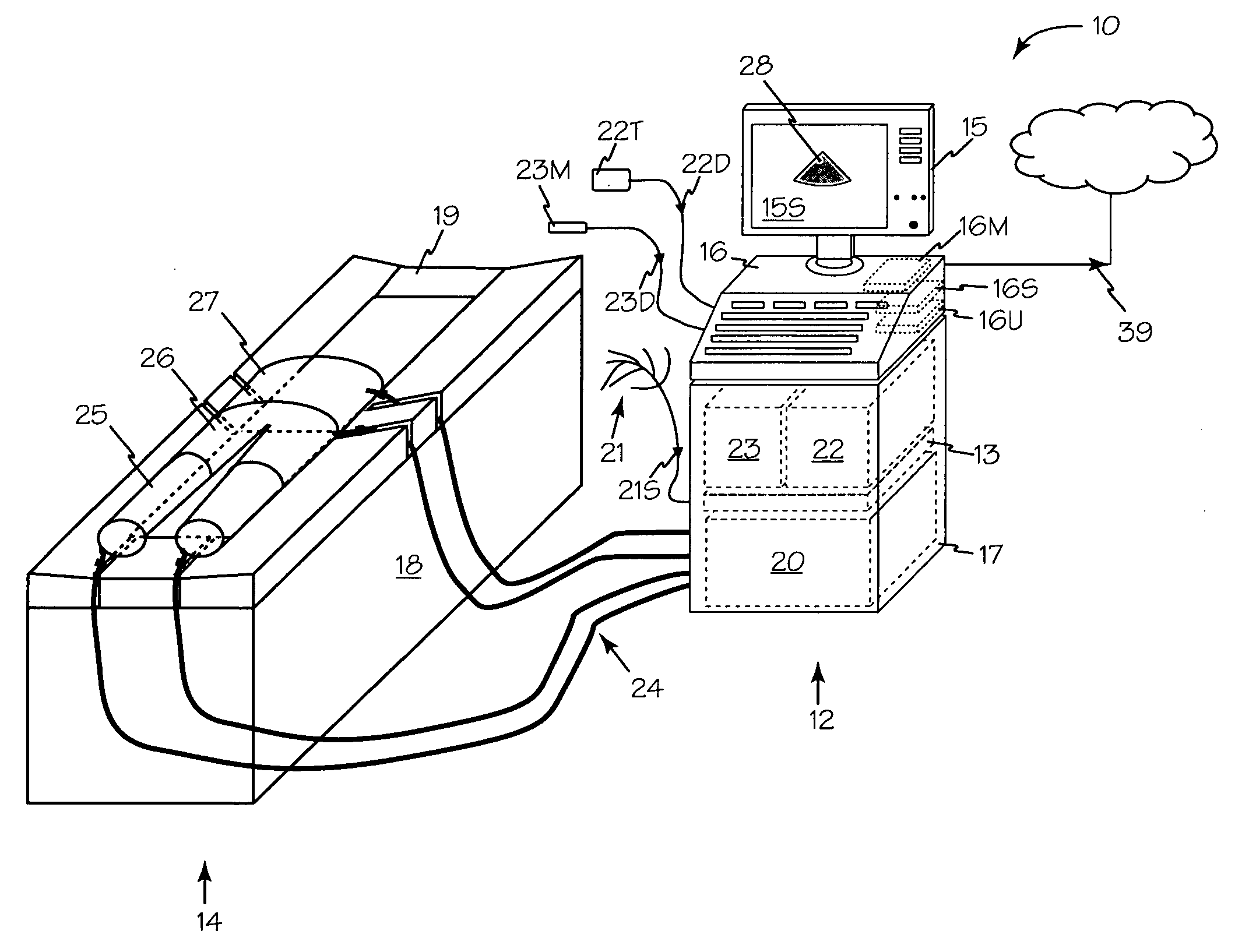
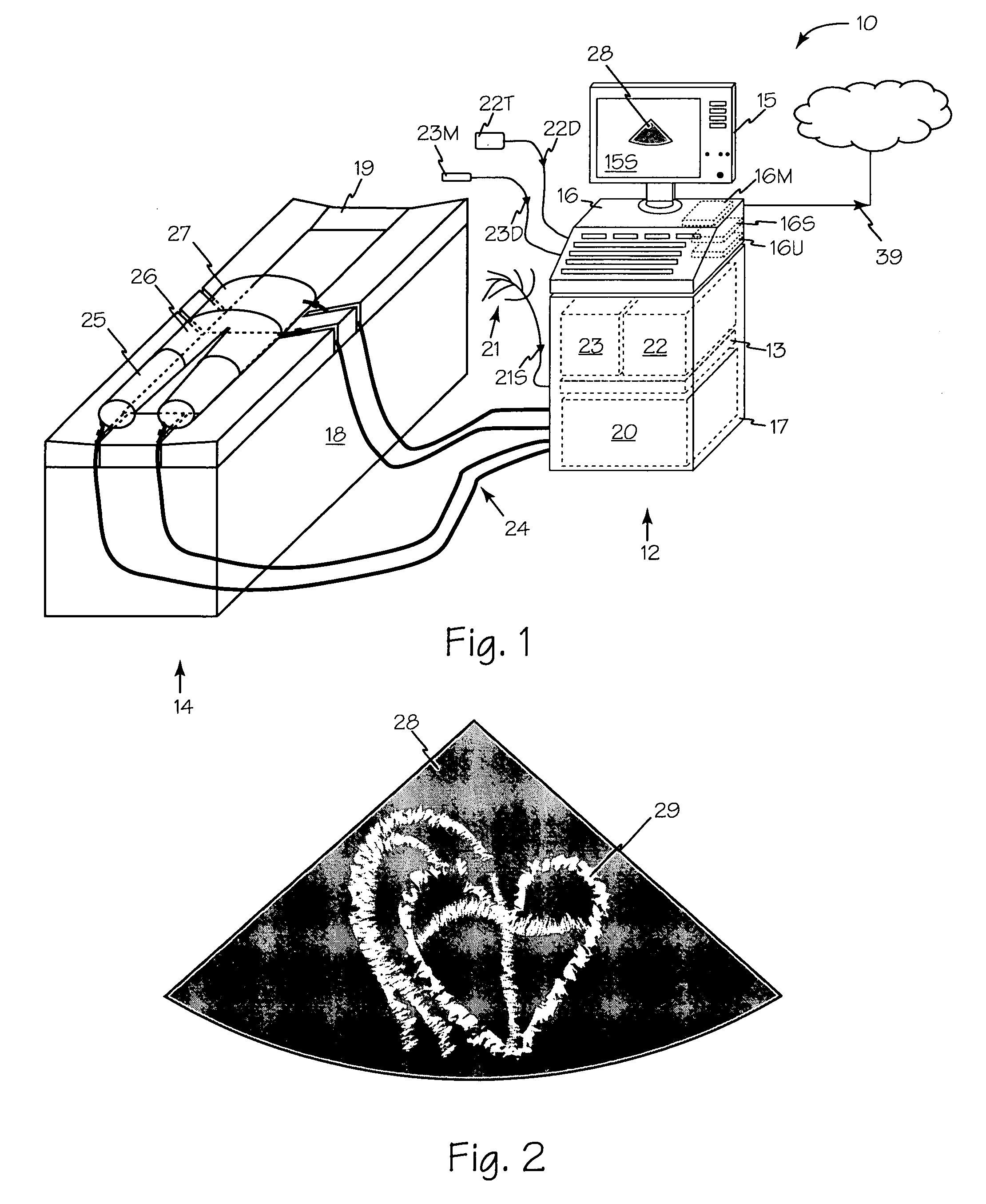
[0018]Image Enhanced External Counterpulsation system 10 of FIG. 1 includes control station 12 and table 14. Control station 12 includes monitor 15, console 16 and enclosure 17. Table 14 includes table base 18 and tabletop or cushion 19.

[0019]Console 16 controls the operation of electrocardiograph (ECG) module 13, ECP driver module 20 and ultrasound driver module 22 and also records data from the ECP driver 20, ECG module 13 and ultrasound system 22 and any other systems or modules that may be combined. ECP driver 20 provides pneumatic pressure through hoses 24 to one or more inflation cuffs such as cuffs 25, 26 and 27. Console 16 may include one or more microprocessors such as microprocessors 16U for processing ultrasound, ECP and any other suitable data.

[0020]Electrocardiograph electrodes 21 are common to both the ECP and ultrasound to ECP timing and ultrasound images are synchronized. One set of electrodes such as ECG electrodes 21 may be used, and electrical signal 21S may be di...


PUM


Abstract

An image enhanced external counterpulsation system may be used to monitor a continuous real-time ultrasound image of the patient's heart during ECP therapy.

Description

RELATED APPLICATIONS[0001]This application claims priority from copending U.S. Provisional patent application 60 / 918,113 filed Mar. 14, 2007.FIELD OF THE INVENTIONS[0002]The inventions described below relate to the field of medicine and more specifically to medical diagnosis and therapy using simultaneous ultrasound, ECG, tonometry and external counterpulsation.BACKGROUND OF THE INVENTIONS[0003]Echocardiography has been used for noninvasive imaging of a heart in operation. Use of this technique has generally been limited to office or laboratory environments with the patient generally immobile (laying, sitting or standing).[0004]Conventional External Counter Pulsation (ECP) treatment usually lasts 35 days with the results of the therapy being subjective, usually based on how the patient describes their current feeling of well being and overall discomfort relative to how they felt prior to the beginning treatment. Furthermore, the only functions monitored continuously during conventio...

Claims


Application Information

Patent Timeline
Patent Type & Authority Applications(United States)
IPC IPC(8): A61M1/10A61B8/00
CPCA61H2230/06A61H9/0078
Inventor MANERA, LOREN A.BURNS, KENNETH L.MALENIUS, WILLIAM R.
Owner RENEW GRP
Why Eureka
Try Eureka