Methods for Isolating Monocytes

a monocyte and anti-mouse technology, applied in the field of monocyte anti-mouse detection antibodies, can solve the problems damage to cells, and achieve the effect of increasing the chance of bacterial contamination

Inactive Publication Date: 2008-10-09
MEDICAL & BIOLOGICAL LAB CO LTD
View PDF1 Cites 11 Cited by
  • Summary
  • Abstract
  • Description
  • Claims
  • Application Information

AI Technical Summary

Benefits of technology

[0014]Dendritic cells and macrophages to be used in cell immunotherapy or such can be induced to differentiate from monocytes. Thus, methods using autologous peripheral blood monocytes separated from patients are in wide clinical use. In methods using peripheral blood monocytes, the monocytes are first separated by apheresis or density centrifugation. Such physical methods require repeated centrifugation, and are problematic in that they significantly damage cells and further increase the chance of bacterial contamination. Some surface antigens, including CD14, CD11b, and CD33, are known as monocyte markers; however, more specific monocyte markers are required. Thus, an objective of the present invention is to identify proteins to become markers that are highly expressed in monocytes.

Problems solved by technology

Such physical methods require repeated centrifugation, and are problematic in that they significantly damage cells and further increase the chance of bacterial contamination.

Method used


Image

  • Methods for Isolating Monocytes
  • Methods for Isolating Monocytes
  • Methods for Isolating Monocytes

Examples


examples

1. Preparation of Anti-HIDE1 Antibodies

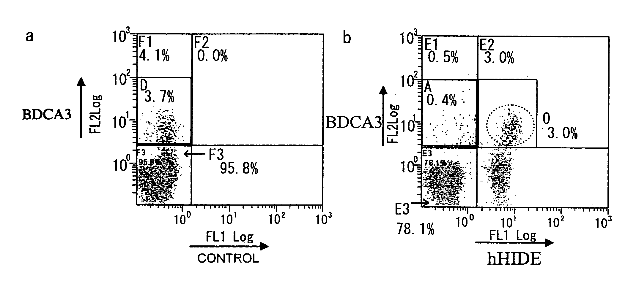
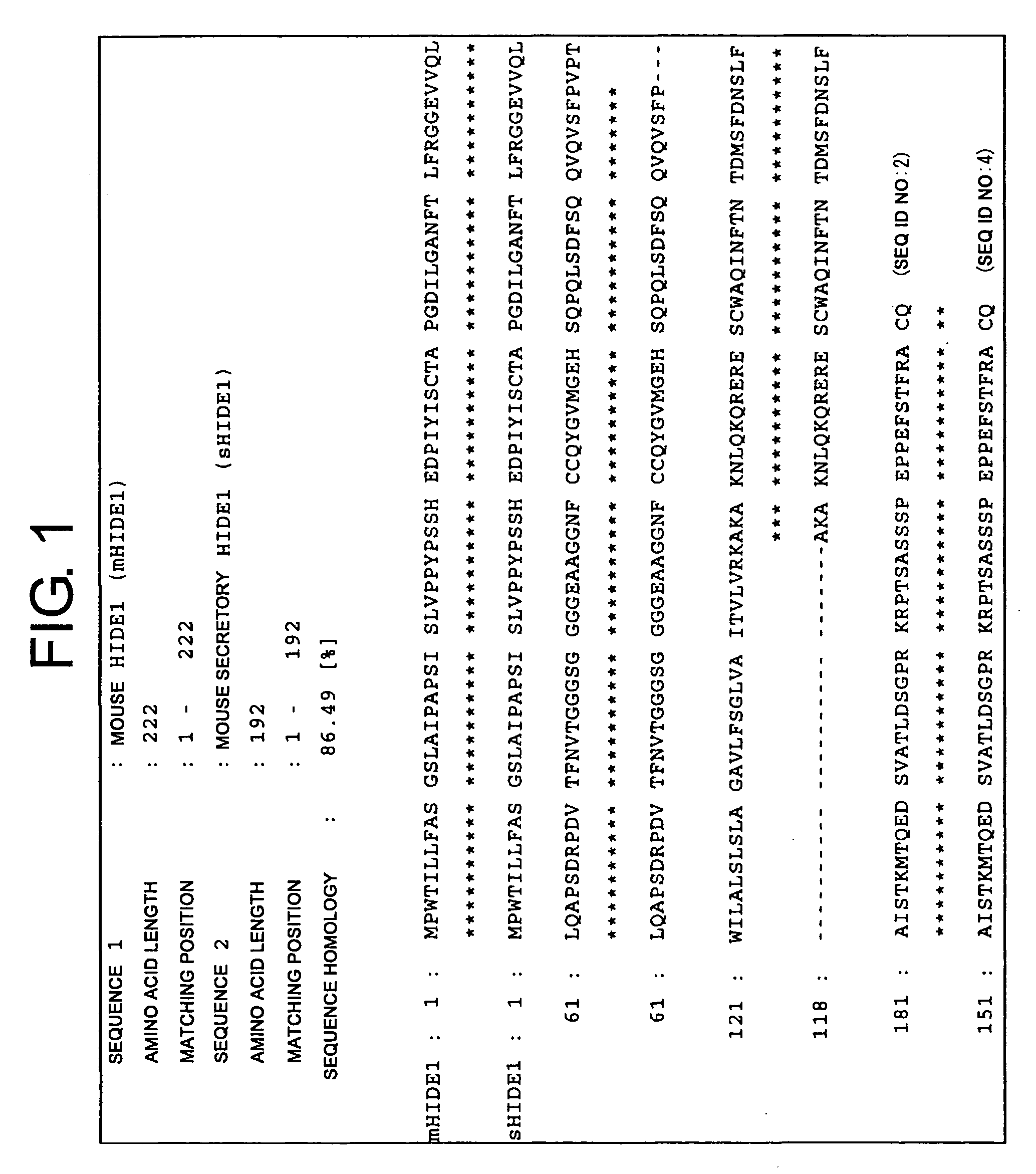
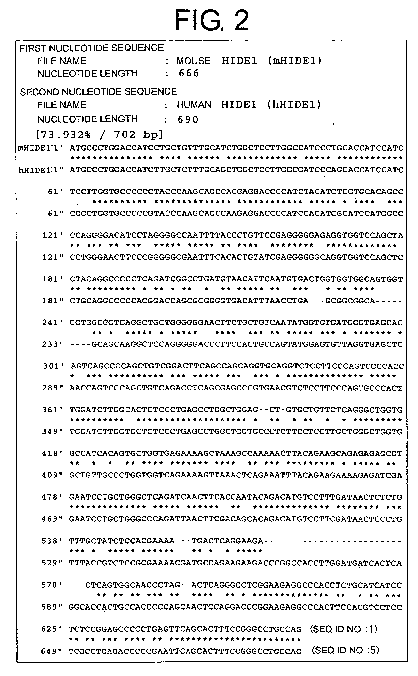
[0172]Monoclonal antibodies against a human homolog of HIDE1 were prepared by the following procedure: First, PCR was used to amplify a human HIDE1 gene from a placental cDNA library. The yielded PCR product was cloned into pcDNA3.1 (Invitrogen), an expression vector for cultured animal cells (pcDNA-hHIDE1). In the constructed vector, the C terminus of human HIDE1 (hHIDE1) is fused with a Myc tag. Cells of the human cultured cell line 293T were transiently transfected with pcDNA-hHIDE1 using Lipofectamine 2000 (Invitrogen), and the yielded cells expressing hHIDE1 were used as antigens to immunize Balb / C mice. Lymphocytes collected from the lymph nodes of the immunized mice were fused with Myeloma P3U1. After ten days, supernatant from each clone cell was collected as samples. Cell supernatants that reacted with the hHIDE1 -expressing cell line were selected by flow cytometry (FCM). Thus, hybridomas producing anti-HIDE1 antibodies were obtained....


PUM

PropertyMeasurementUnit
volumeaaaaaaaaaa
molecular weightaaaaaaaaaa
pHaaaaaaaaaa

Abstract

The present invention provides HIDE1 as novel monocyte markers. Since HIDE1 are membrane proteins, monocytes can be specifically detected by using antibodies that bind to HIDE1. Further, HIDE1-positive monocytes can also be collected from peripheral blood or the like using a cell sorter, magnet, or such. Monocytes that can be prepared based on the present invention are useful in cell immunotherapy.

Description

[0001]This application is a U.S. National Phase Application, filed under 35 U.S.C. §371 of Patent Cooperation Treaty Application No. PCT / JP2005 / 00923, filed Jan. 25, 2005 and claims the benefit of Japanese Patent Application Number 2004-018747, filed Jan. 27, 2004, the contents of each of the aforementioned applications are hereby incorporated by reference in their entirety.TECHNICAL FIELD[0002]The present invention relates to antibodies for detecting monocyte markers, and uses thereof.BACKGROUND ART[0003]Monocytes are cells which migrate in the blood and which have phagocytotic activity, belonging to the group of mononuclear phagocytes. Once differentiated, monocytes remain in the bone marrow for only short time before entering the circulatory system, where they remain for several days. Monocytes then infiltrate tissues and body cavities, and differentiate into macrophages and dendritic cells. Monocytes are known to increase in the circulatory system in inflammatory diseases, post-...

Claims


Application Information

Patent Timeline
Patent Type & Authority Applications(United States)
IPC IPC(8): A61K35/12C07K16/18G01N33/569A61P43/00A01N1/02C12N5/06C07K16/28C12N5/0786
CPCA61K2035/124C07K16/2803C12N5/0645G01N33/56972A61P25/00A61P31/00A61P35/00A61P37/00A61P37/04A61P37/06A61P43/00
Inventor NAKAGAWA, TOMOKOKUREI, SHUNSUKETOJI, SHINGOOKABE, AYAKOKUHARA, MOTOKIKISHI, YOSHIROYAHARA, ICHIRO
Owner MEDICAL & BIOLOGICAL LAB CO LTD
Why Eureka
Try Eureka