Cerebral Perfusion Monitor

a perfusion monitor and cerebral technology, applied in the field of measuring blood flow in the head, can solve the problems of increased blood flow to the brain, brain cell death, brain cell damage, etc., and achieve the effect of poor ability to maintain constant blood flow, and rapid initial rise of ipg signal

Inactive Publication Date: 2008-11-06
ORSAN MEDICAL TECH
View PDF41 Cites 25 Cited by
  • Summary
  • Abstract
  • Description
  • Claims
  • Application Information

AI Technical Summary

Benefits of technology

[0015]An aspect of some embodiments of the invention relates to estimating cerebral blood flow by using characteristics of the IPG signal alone. For example, the cerebral blood flow is estimated from the peak value of the IPG signal in each cardiac cycle, or from the peak rate of rise of the IPG signal after the beginning of each cardiac cycle, or from the height of the first local peak or inflection point in the IPG signal after the beginning of each cardiac cycle. The beginning of each cardiac cycle is defined, for example, by the peak of the R-wave of an ECG, or by the time of the minimum in the IPG or PPG signal, or by the time of the diastolic pressure. The rapid initial rate of rise in the IPG signal, up to the peak or up to the first local peak or inflection point, may be dominated by the cerebral blood flow, even if the IPG signal during the rest of the cardiac cycle is largely influenced by the scalp blood volume, since the scalp blood volume, as indicated by PPG data, generally rises more slowly, and with a delay, at the beginning of each cardiac cycle. Optionally, PPG data is also obtained, to confirm that the scalp blood volume is rising slowly initially, and that the rapid initial rise of the IPG signal is indeed due mostly to the cerebral blood flow.
[0016]Some embodiments of the invention may be particularly useful for monitoring premature infants, for example those with weight under 1.5 kg, who generally have poor ability to maintain constant blood flow to the brain due to the immaturity of their cerebral blood flow autoregulation system. Abrupt changes in blood flow to the brain can be caused by changes in respiration, changes in blood pressure, and manipulation of the infants by medical staff. Such abrupt changes in cerebral blood flow, if not immediately detected and treated, can cause severe brain injury, including injuries caused by cerebral hemorrhage which occurs in 10% to 30% of premature babies. The invention may also be useful in monitoring mature babies who may be at risk of brain hemorrhage or ischemia for various reasons.
[0017]The invention may also be useful for monitoring cerebral blood flow in 1) patients undergoing surgery of the carotid arteries, in which a clamp is applied to one of the carotids, potentially reducing blood flow to the brain; 2) patients with stenosis or occlusion of the carotid arteries or cerebral arteries, particularly if they are undergoing procedures such as intra-arterial catherization or stent application in the affected arteries; 3) brain injury patients, in whom brain edema might cause a decrease in blood perfusion, and herniation of the brain; 4) neurosurgery patients, during and for a few days after the surgery, when cerebral blood flow is often impaired; 5) patients undergoing other major surgery, including heart surgery, in which massive bleeding and resulting hypotension could lead to a decrease in cerebral blood flow. In all of these categories of patients, monitoring of cerebral blood flow could lead to prompt intervention before brain injury occurs.

Problems solved by technology

There is a need to measure cerebral blood flow during various medical events and procedures, because any disturbance to the flow of blood to the brain may cause injury to the function of the brain cells, and even death of brain cells if the disturbance is prolonged.
A sudden increase in blood flow to the brain may also cause severe damage, and is especially likely to occur in newborn or premature babies, although such an increase may also occur in other patients with certain medical conditions, or during surgery.
But due to the different relation between blood pressure and cerebral blood flow in different medical conditions, there may be situations in which cerebral blood flow is inadequate even when blood pressure appears to be adequate.
Existing means for measuring cerebral blood flow are complex, expensive, and in some cases invasive, which limits their usefulness.
Three methods that are presently used only in research are 1) injecting radioactive xenon into the cervical carotid arteries and observing the radiation it emits as it spreads throughout the brain; 2) positron emission tomography, also based on the injection of radioactive material; and 3) magnetic resonance angiography, performed using a room-sized, expensive, magnetic resonance imaging system, and requiring several minutes to give results.
These three methods can only be carried out in a hospital or other center that has the specialized equipment available, and even in a hospital setting it is not practical to monitor patients continuously using these methods.
However, TCD fails to give a correct determination of blood flow in about 15% of patients, due to the difficulty of passing sound waves through the cranium, and it requires great skill by professionals who have undergone prolonged training and practice in performing the test and deciphering the results.
Another disadvantage of TCD is that it measures only regional blood flow in the brain, and does not measure global blood flow.

Method used


Image

  • Cerebral Perfusion Monitor
  • Cerebral Perfusion Monitor
  • Cerebral Perfusion Monitor

Examples


Embodiment Construction

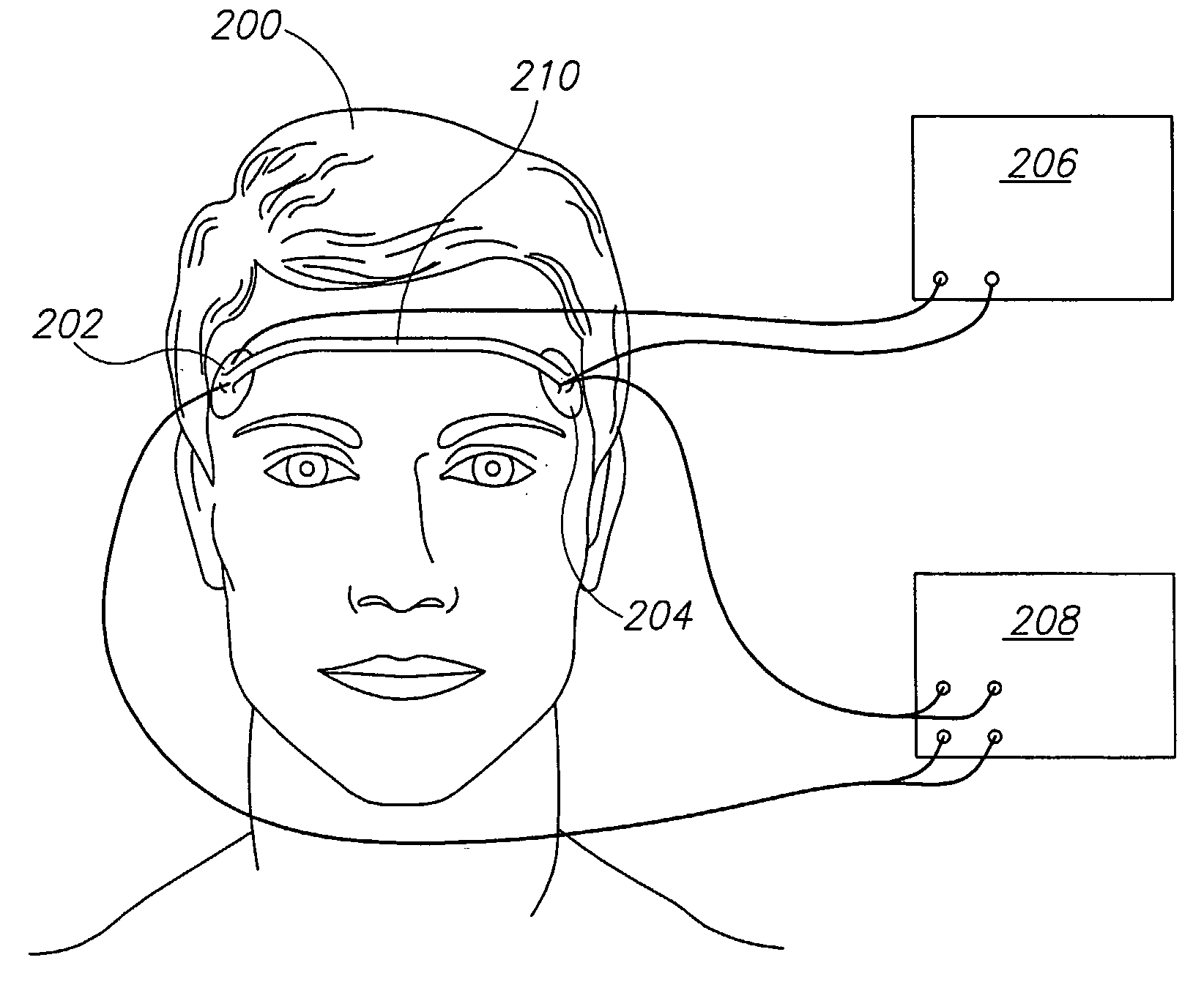
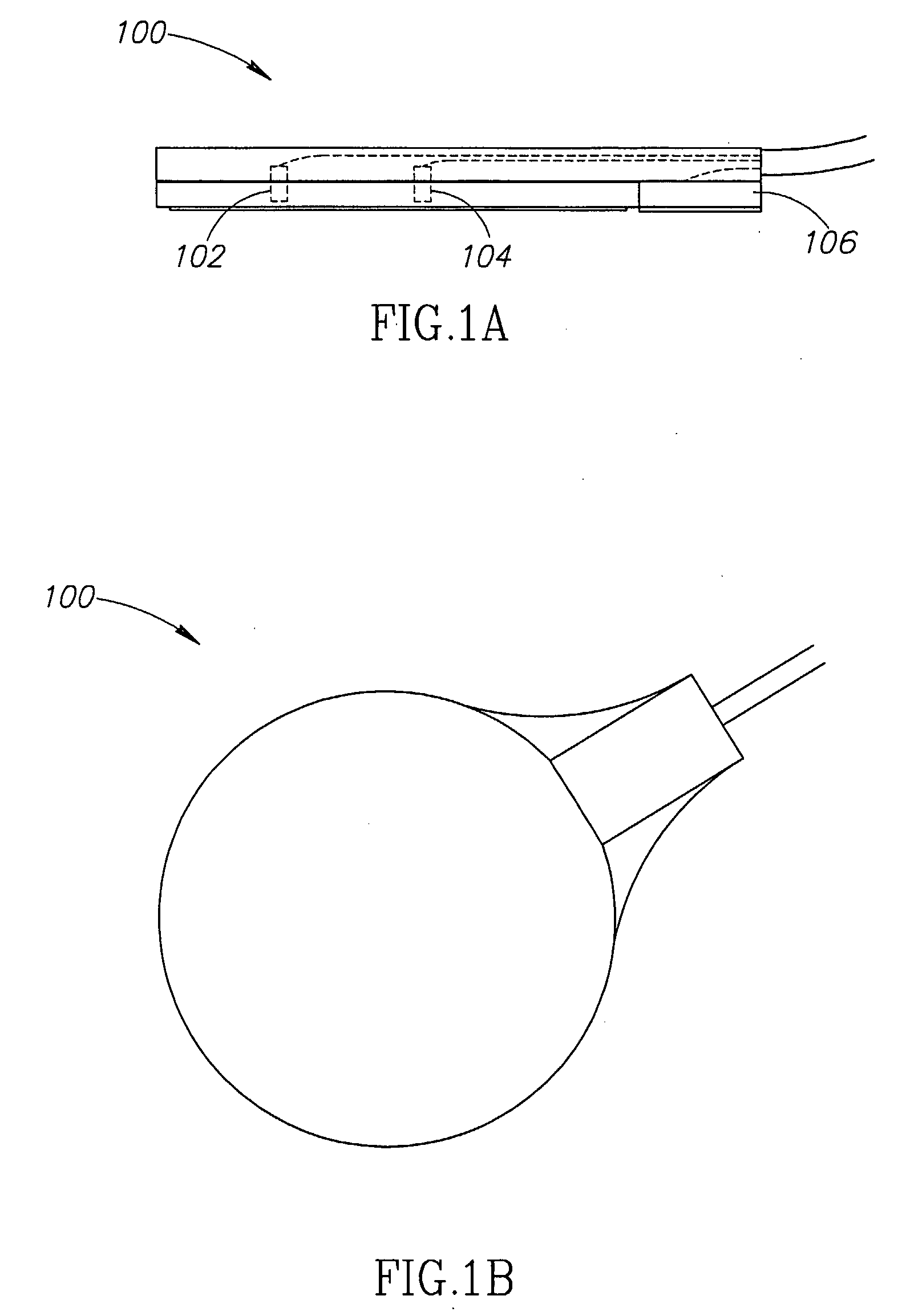
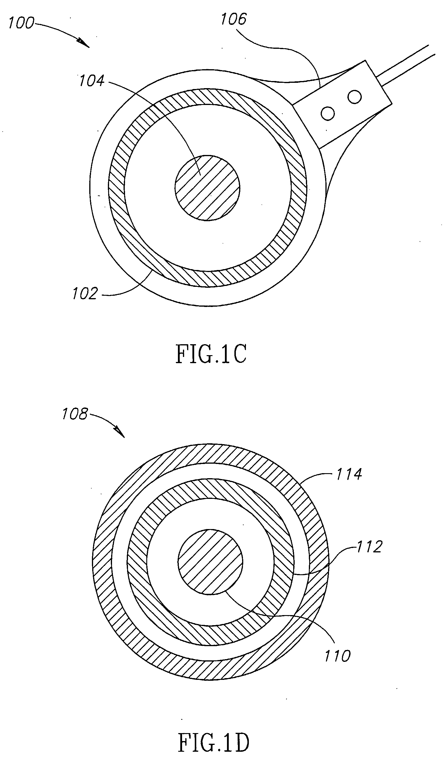
[0066]FIGS. 1A, 1B, and 1C respectively show side, back, and face views of a unit 100 which optionally combines a current electrode 102 and a voltage electrode 104 for impedance plethysmography (IPG), and a sensor 106 for photoplethysmography (PPG), according to an exemplary embodiment of the invention. The face side of unit 100, shown in FIG. 1C, is the side that is placed against the skin, as shown in FIG. 2. As shown in FIG. 2, two such units, placed for example on opposite sides of the head, are optionally used for IPG, passing current from one unit to the other and measuring the voltage between them. For reasons described below, alternating current is generally used.

[0067]PPG sensor 106 measures the color of the skin to determine a degree of perfusion of oxygenated blood in the skin adjacent to unit 100, as described, for example, by J. Webster, “Measurement of Flow and Volume of Blood,” in John G. Webster (ed.), Medical Instrumentation: Application and Design (Wiley, 1997), th...


PUM


Abstract

A method of estimating cerebral blood flow includes obtaining a measure of time-varying blood volume in the head, using an impedance plethysmography (102 and 104), obtaining a measure of time-varying blood volume in the scalp, and using the time-varying blood volume in the head and scalp to estimate cerebral blood flow.

Description

RELATED APPLICATIONS[0001]The present application is a continuation-in-part of U.S. patent application Ser. No. 10 / 893,570, filed Jul. 15, 2004, which is a continuation-in-part of PCT patent application PCT / IL03 / 00042, filed Jan. 15, 2003, which claims benefit under 35 USC 119(e) from U.S. provisional patent application 60 / 348,278, filed Jan. 15, 2002. This application is related to a PCT application filed on even date, titled “Device For Monitoring Blood Flow To Brain,” having attorney docket number 371 / 04609. The disclosures of all of these applications are incorporated herein by reference.FIELD OF THE INVENTION[0002]The field of the invention relates to measuring blood flow in the head.BACKGROUND OF THE INVENTION[0003]There is a need to measure cerebral blood flow during various medical events and procedures, because any disturbance to the flow of blood to the brain may cause injury to the function of the brain cells, and even death of brain cells if the disturbance is prolonged....

Claims


Application Information

Patent Timeline
Patent Type & Authority Applications(United States)
IPC IPC(8): A61B5/02A61B5/026A61B5/0265A61B5/296
CPCA61B5/0261A61B5/0265A61B5/04008A61B5/0535A61B5/6814A61B5/6817A61B2562/164A61B5/0295A61B5/245
Inventor SHAPIRA, AHARONRAPPAPORT, ALONBEN-ARI, SHLOMIREICHMAN, YOSEFBARNEA, OFER
Owner ORSAN MEDICAL TECH
Why Eureka
Try Eureka