Auto-Alignment and Auto-Focus System and Method

a technology of autofocus and alignment, applied in the field of autoalignment and autofocus system and method, can solve problems such as difficulty in alignment, achieve the effects of improving patient alignment, enhancing speed, ease, safety and efficacy, and increasing or maximizing gradients

Active Publication Date: 2008-11-13
AMO DEVMENT +1
View PDF22 Cites 77 Cited by
  • Summary
  • Abstract
  • Description
  • Claims
  • Application Information

AI Technical Summary

Benefits of technology

[0006]The present invention generally provides improved devices, systems, and methods for supporting and positioning patients. Embodiments of the present invention provide an improved patient alignment between a support structure, such as a chin rest, chair, bed, or table, and a diagnostic instrument, such as a wavefront measurement device, in which the instrument can be moved into alignment with the patient. Other embodiments provide mechanisms for positioning the head and body of a patient to align the patient with an instrument during surgery. Embodiments of the present invention may be particularly useful for enhancing the speed, ease, safety, and efficacy of diagnostic eye measurements and laser eye surgical procedures such as photorefractive keratectomy (“PRK”), laser in situ keratomileusis (“LASIK”), and the like. Embodiments of a patient positioning system may include an image sensor to capture an image of a tissue structure of the eye, for example the boundary between the iris and pupil of the eye. A linkage may be coupled to a patient support and an image sensor support, such that the linkage can move the patient support and / or the image sensor support to align the image sensor with the patient. Gradients and / or slopes of illumination levels of the tissue structure can be determined, for example calculated with a processor. A separation distance from the image sensor to the tissue structure can be adjusted in response to the gradients and or slopes of the tissue structure in the image so as to bring the image of the tissue structure into focus. In some embodiments, the separation distance is adjusted so as to increase or maximize peaks of the gradients and / or to reduce or minimize widths of edges in the tissue structure image. A location of the pupil and / or iris in the image may be determined, and the linkage driven to align the pupil and / or iris with a predetermined location, for example a central position in the image.
[0015]In some embodiments, the linkage can move the patient support to adjust the separation distance in response to the gradient of the tissue structure image.

Problems solved by technology

In some instances, the patient may move while the patient is supported, such that alignment can be difficult in some instances.

Method used


Image

  • Auto-Alignment and Auto-Focus System and Method
  • Auto-Alignment and Auto-Focus System and Method
  • Auto-Alignment and Auto-Focus System and Method

Examples


Embodiment Construction

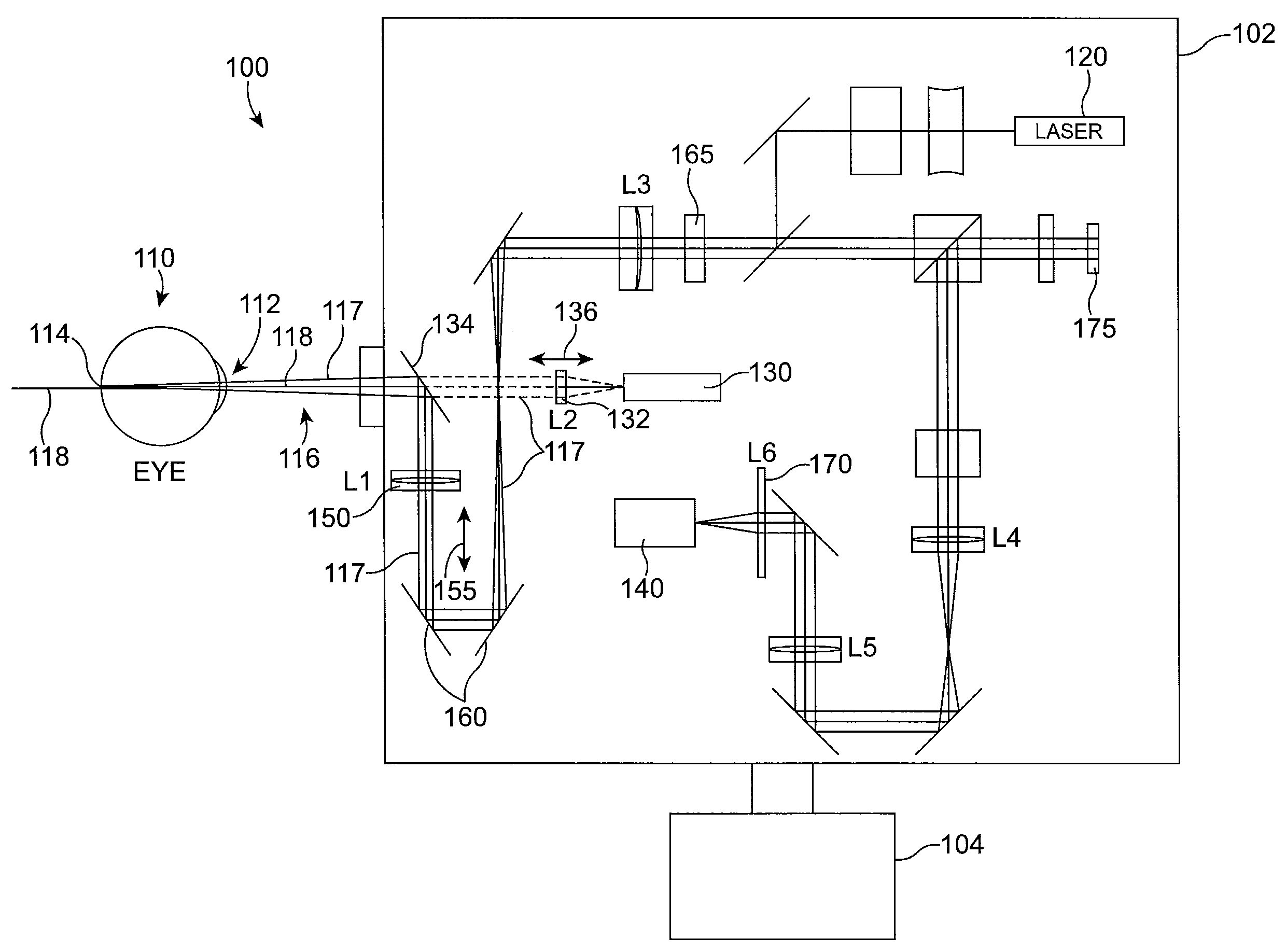
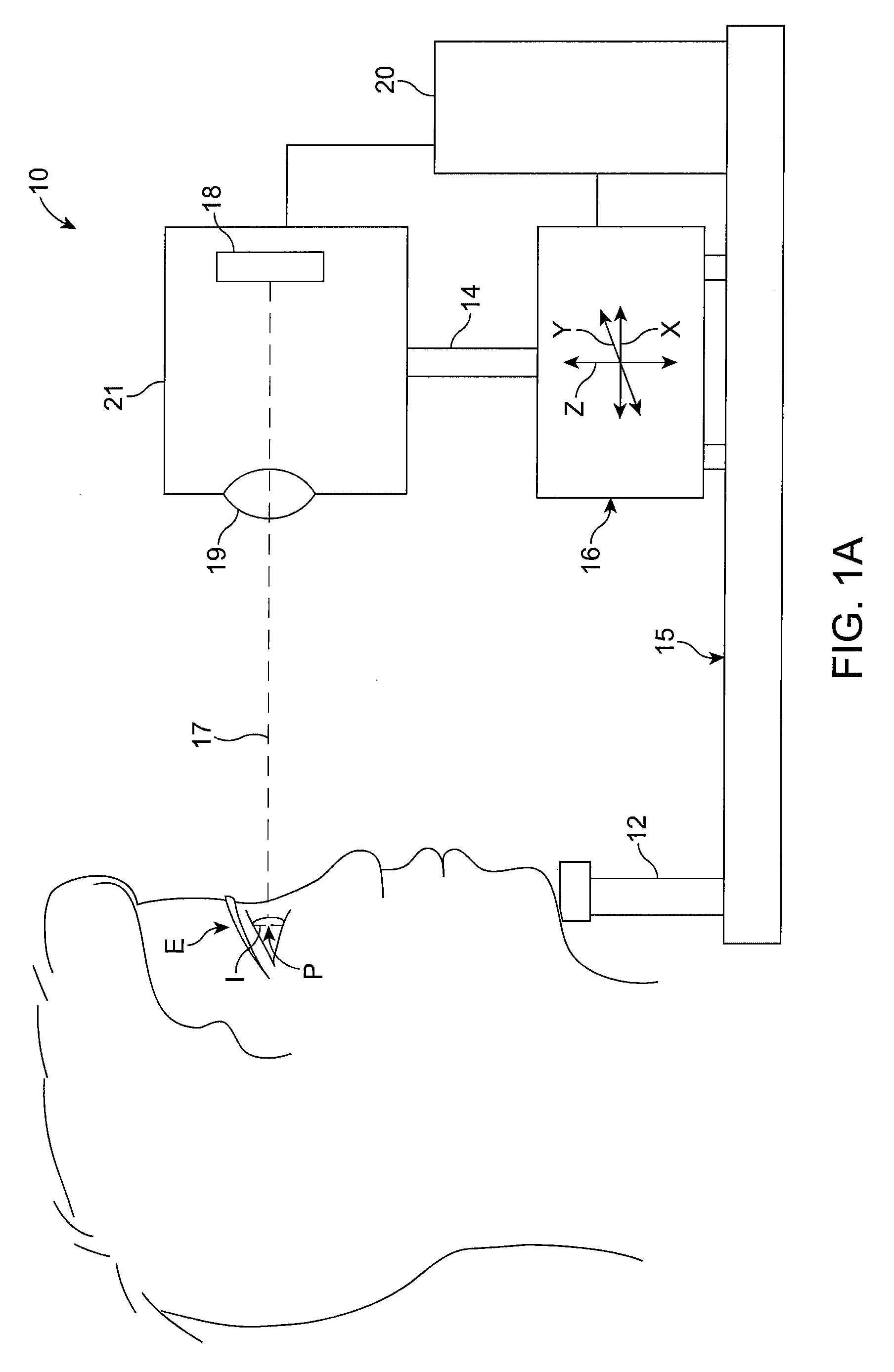
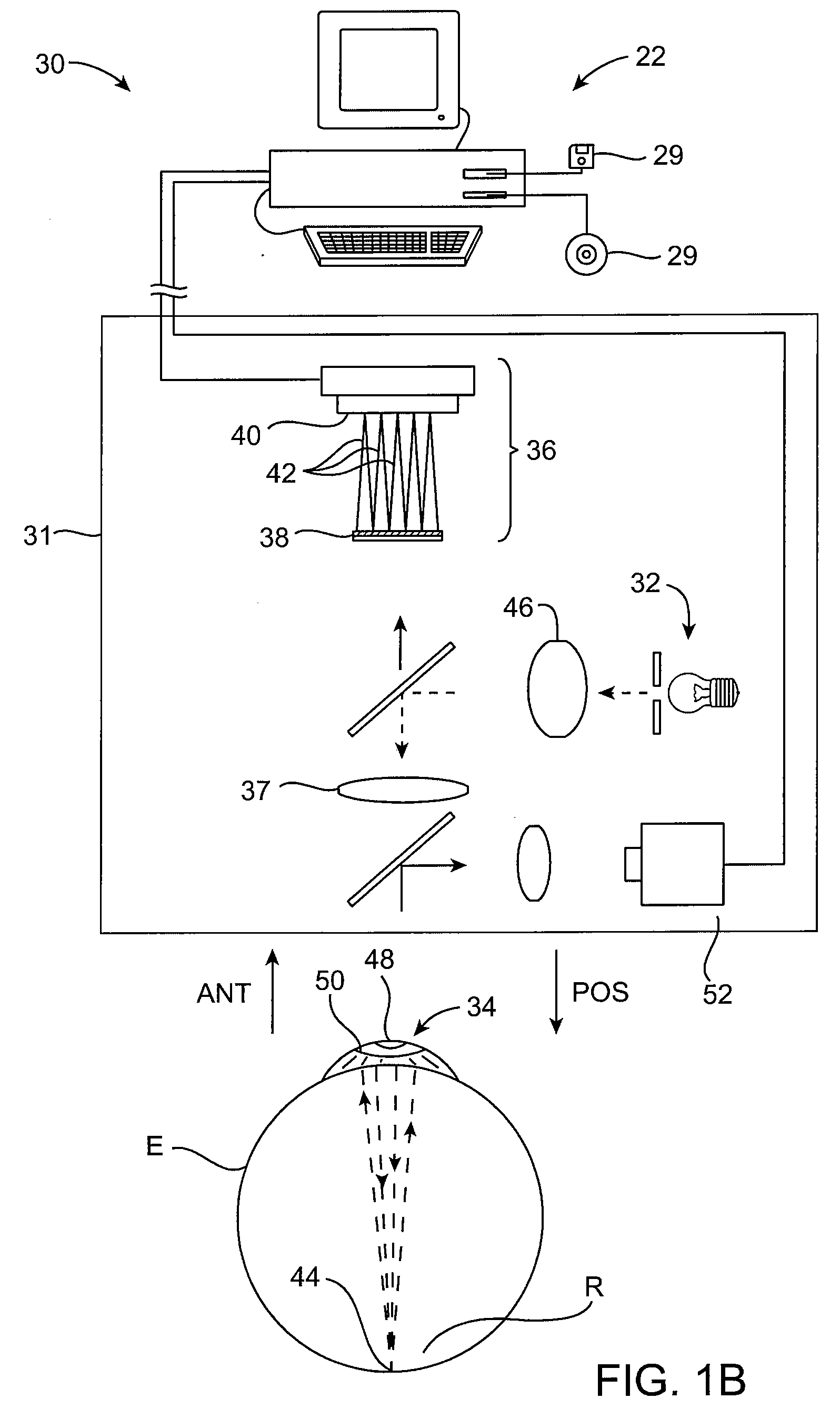
[0037]The present invention relates generally to devices, systems, and methods for supporting and positioning patients and / or for analyzing ocular images. Embodiments of the present invention provide an improved patient alignment between a support structure, such as a chin rest, chair, bed, or table, and a diagnostic instrument, such as a wavefront measurement device, in which the instrument can be moved into alignment with the patient. Other embodiments provide mechanisms for positioning the head and body of a patient and stabilizing the patient support, providing improved patient stability during surgery. Although specific reference is made to images of ocular tissue structures comprising an iris and a pupil of the eye, embodiments of the invention may used to image and focus on other ocular tissue structures, for example the limbus of the eye. Embodiments of the present invention may be particularly useful for enhancing the speed, ease, safety, and efficacy of diagnostic eye meas...


PUM


Abstract

A patient positioning system for use with a patient comprises an optical image sensor. The optical image sensor measures an optical image of a tissue structure of an eye of the patient. An image sensor support supports the image sensor. A patient support supports the patient. A linkage is coupled to the image sensor support with the patient support. A processor is coupled to the optical image sensor to determine a gradient of an illumination level of the optical tissue structure image. The processor is coupled to the linkage and configured to articulate the linkage so as to adjust a separation distance from the tissue structure to the image sensor in response to the gradient of the optical tissue structure image.

Description

BACKGROUND OF THE INVENTION[0001]The present invention relates generally to devices, systems, and methods for supporting and aligning patients with instruments and / or for analyzing ocular images. Exemplary embodiments of the present invention provide patient alignment between a support structure, such as a chin rest, chair, bed, or table, and a diagnostic instrument, such as a wavefront measurement device, in which the instrument can be moved into alignment with the patient. Other embodiments provide mechanisms for positioning the head and / or body of a patient to align the patient with an instrument during surgery.[0002]Laser eye surgical procedures typically employ ultraviolet or infrared lasers to remove a microscopic layer of stromal tissue from the cornea to alter the cornea's refractive properties. Excimer laser systems generally use argon and fluorine gas to create a non-thermal laser light which can break molecular bonds in a process known as photoablation. Such systems resul...

Claims


Application Information

Patent Timeline
Patent Type & Authority Applications(United States)
IPC IPC(8): A61B3/15
CPCA61B3/0075A61B3/0083A61F2009/00872A61F2009/00846A61F2009/00848A61F9/00806
Inventor SOMANI, SEEMA
Owner AMO DEVMENT
Why Eureka
Try Eureka