Fusion Proteins That Bind Effector Lymphocytes And Target Cells
a technology of effector lymphocytes and fusion proteins, applied in the field of proteins, can solve the problems of limited flexibility of such constructs and laborious generation of bispecific antibodies composed entirely of human antibody sequences
- Summary
- Abstract
- Description
- Claims
- Application Information
AI Technical Summary
Benefits of technology
Problems solved by technology
Method used
Image
Examples
example 1
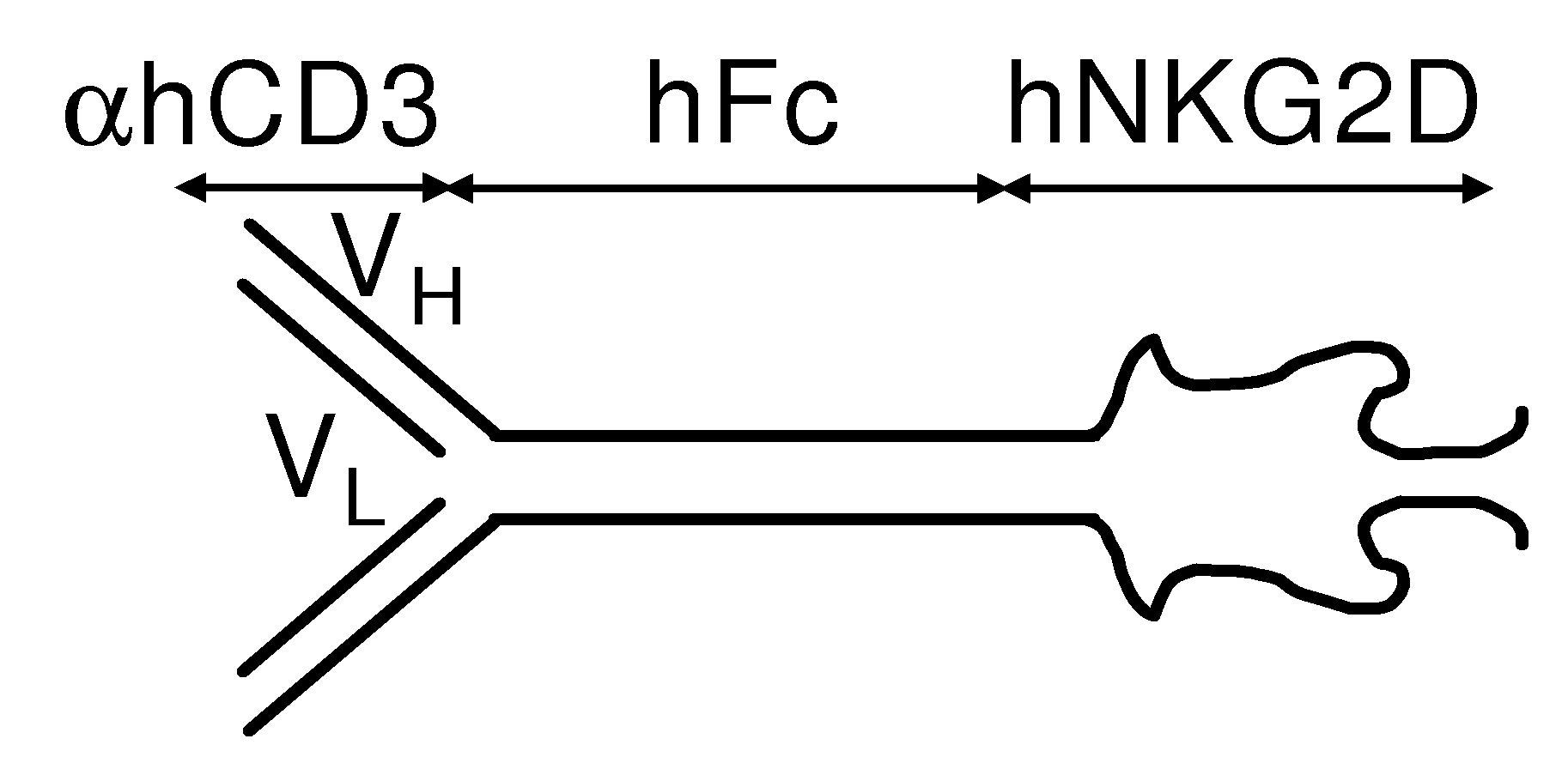
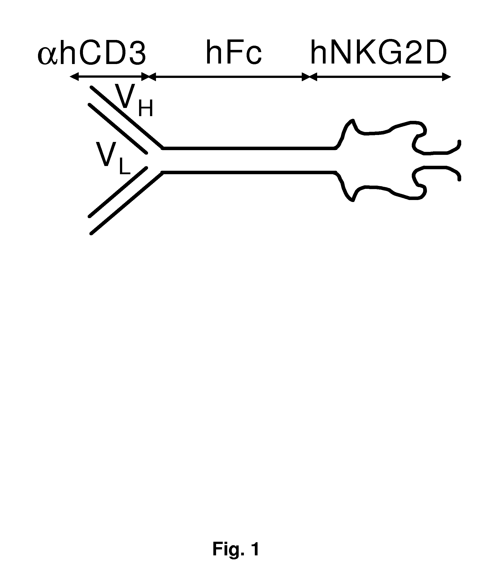
[0222]The following example describes the production of a fusion protein comprising a anti-aCD3 portion and an NKG2D portion.
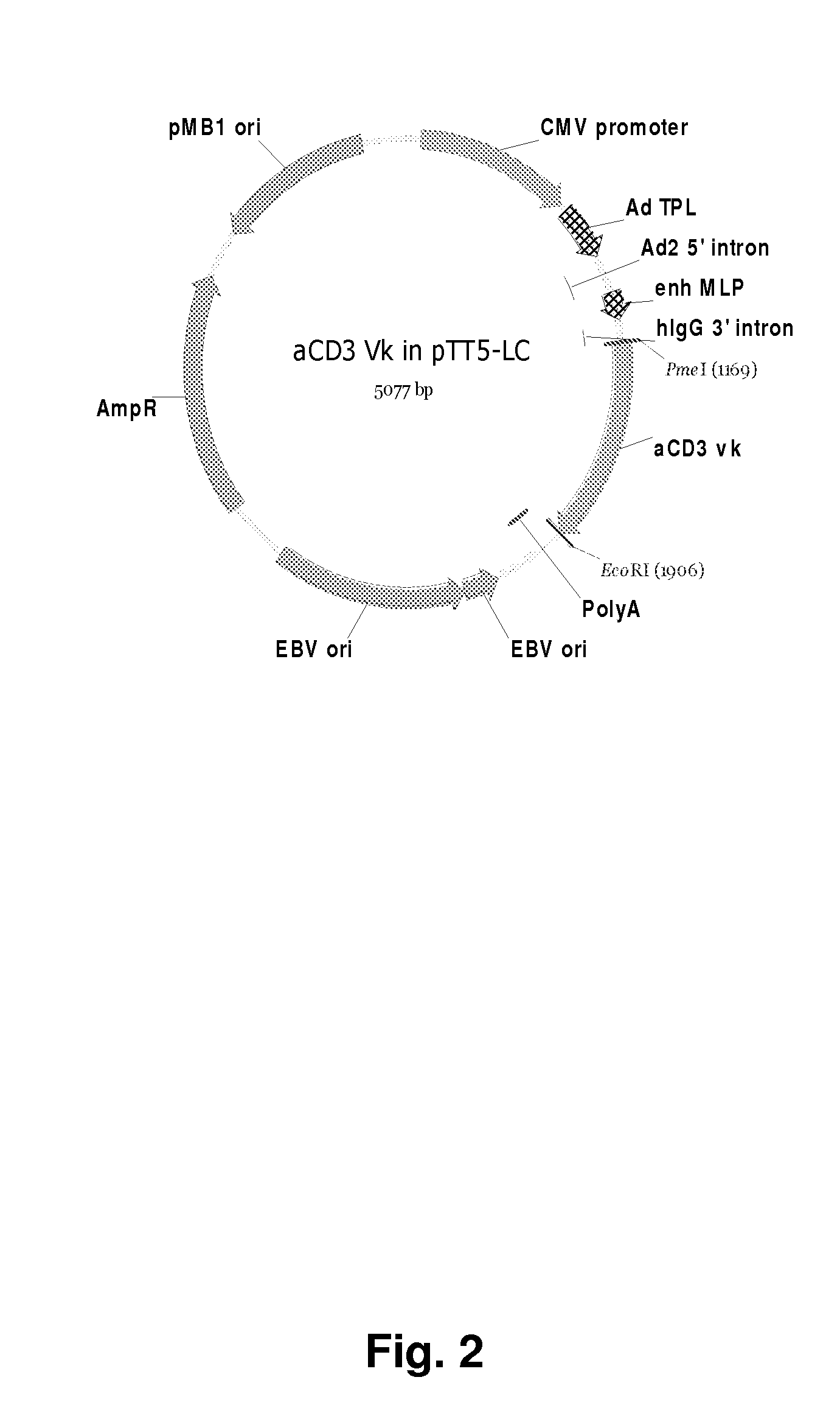
[0223]cDNA encoding an anti-mouse CD3 antibody was cloned from a hamster anti-mouse CD3 producing cell line (145-2c11). The total RNA was purified according to manufacturers instructions (RNeasy from Qiagen, VWR, Denmark) and the gene sequences amplified using specific primers in a RT-PCR reaction using SuperScript™ III One-Step RT-PCR System with Platinum® Taq DNA Polymerase from Invitrogen and the reaction cycles: [37° C. 30 min][94° C.] min] 25×[94° C. 30 s; 55° C. 30 s; 72° C. min][72° C. 5 min]. The RT-PCR products were analyzed by electrophoresis on a 1% agarose gel and the DNA purified from the gel using GFX PCR and Gel Band Purification Kit (Amersham Biosciences, Denmark). The primers used to obtain the anti-mouse CD3 light chain were oligonucleotides oVWS109 and oVWS110.
oVWS109 (145.2c11 light forward) (SEQ ID NO:22):5′ ATGAGGGCCCCTACTGTGTATCC 3′oVWS1...
example 2
[0240]This example demonstrates a method by which the functionality of an exemplary fusion protein of the invention may be assessed.
[0241]The functionality of the fusion protein produced in Example 1 was assessed in vitro by Cr51 release assay. Cells carrying a NKG3D ligand such as MicA (e.g., HEK293 cells) were used as target cells and cytotoxic T cells that do not express a NKG2D ligand as effector cells. The target cells are incubated in a solution containing a radioactive isotope of chromium, chromium 51 (Cr51). Cr51 is spontaneously taken up into the cells and stored in the cytosol, and excess chromium containing solution is washed away. Activated CD8 cells are then added to the cell-containing media, and both cell types are incubated together.
[0242]During this period of co-incubation, the activated CD8 lymphocytes will eventually recognize the target cells and cause cell lysis. As the cells lyse, the chromium that they had taken up is released into the supernatant of the mixtu...
example 3
[0244]This example describes how to produce a fusion protein comprising a human anti-αCD3 portion and a human NKG2D portion.
[0245]In order to make the fusion protein, total RNA is purified according to manufacturers instructions (RNeasy from Qiagen, VWR, Denmark) from a hybridoma cell line, OKT3, expressing the mouse monoclonal antibody against human T cell CD3.
[0246]The cDNAs of the variable heavy (VH) and variable light (VL) chains are amplified by polymerase chain reaction (PCR) method using the SMART RACE (Rapid Amplification of cDNA Ends) cDNA Amplification Kit from Clontech (BD Bioscience, Denmark) according to manufacturers instructions using 1 μg of the purified RNA (described above), the 5′ RACE CDS primer and BD SMART II A oligo.
5′ RACE CDS primer:5′ (T)25VN 3′ (V = A,G, og C; N = A,C,G, or T)BD SMART II A oligo (SEQ ID NO:39):5′ AAGCAGTGGTATCAACGCAGAGTACGCGGG 3′
[0247]The VH and VL regions of OKT3 cDNA (made as described above) are amplified by PCR according to the manufac...
PUM
| Property | Measurement | Unit |
|---|---|---|
| molecular weight | aaaaa | aaaaa |
| molecular weight | aaaaa | aaaaa |
| time | aaaaa | aaaaa |
Abstract
Description
Claims
Application Information
 - R&D
- Intellectual Property
- Life Sciences
- Materials
- Tech Scout
- Unparalleled Data Quality
- Higher Quality Content
- 60% Fewer Hallucinations
Browse by: Latest US Patents, China's latest patents, Technical Efficacy Thesaurus, Application Domain, Technology Topic, Popular Technical Reports.
© 2025 PatSnap. All rights reserved.Legal|Privacy policy|Modern Slavery Act Transparency Statement|Sitemap|About US| Contact US: help@patsnap.com


