
Method for Employing a Biosensor to Detect Small Molecules ("Fragments") that Bind Directly to Immobilized Protein Targets

a biosensor and fragment technology, applied in the direction of material excitation, instruments, measurement devices, etc., can solve the problems of weak binding, difficult to generate novel leads, complex identification of high-quality lead compounds for druggable protein targets, etc., to achieve less reagents, less time for determination, and more economical

Inactive Publication Date: 2008-12-04
X BODY
View PDF99 Cites 33 Cited by
  • Summary
  • Abstract
  • Description
  • Claims
  • Application Information

AI Technical Summary

Benefits of technology

[0007]Therefore, the invention provides for the determination of very low molecular weight compounds, known to one familiar with the art as fragments, binding to target biomolecules. The invention provides the rigorousness of typical biochemical tests for quantifying and qualifying the interaction as well as providing for the determination of other properties of the fragment molecule. The methods are much more economical, requiring far less time for the determination and requiring much less reagent than one typically finds employed for the same determination using traditional methods such as NMR spectroscopy, x-ray crystallography, or calorimetry.

Problems solved by technology

Identification of high quality lead compounds for druggable protein targets is a complex task.
It can be difficult to generate leads that are novel and optimizable.
Even though low molecular weight compounds can lead to development of efficacious drugs, identification of weak binding, very low molecular weight compounds is extremely slow and difficult.
Prior identification methods for identifying fragment compounds that bind to proteins involve considerable amounts of protein, are labor intensive, require expensive instrumentation operated by highly educated individuals, and take many days or weeks to screen a few small compounds.

Method used


Image

  • Method for Employing a Biosensor to Detect Small Molecules ("Fragments") that Bind Directly to Immobilized Protein Targets
  • Method for Employing a Biosensor to Detect Small Molecules ("Fragments") that Bind Directly to Immobilized Protein Targets
  • Method for Employing a Biosensor to Detect Small Molecules ("Fragments") that Bind Directly to Immobilized Protein Targets

Examples


examples

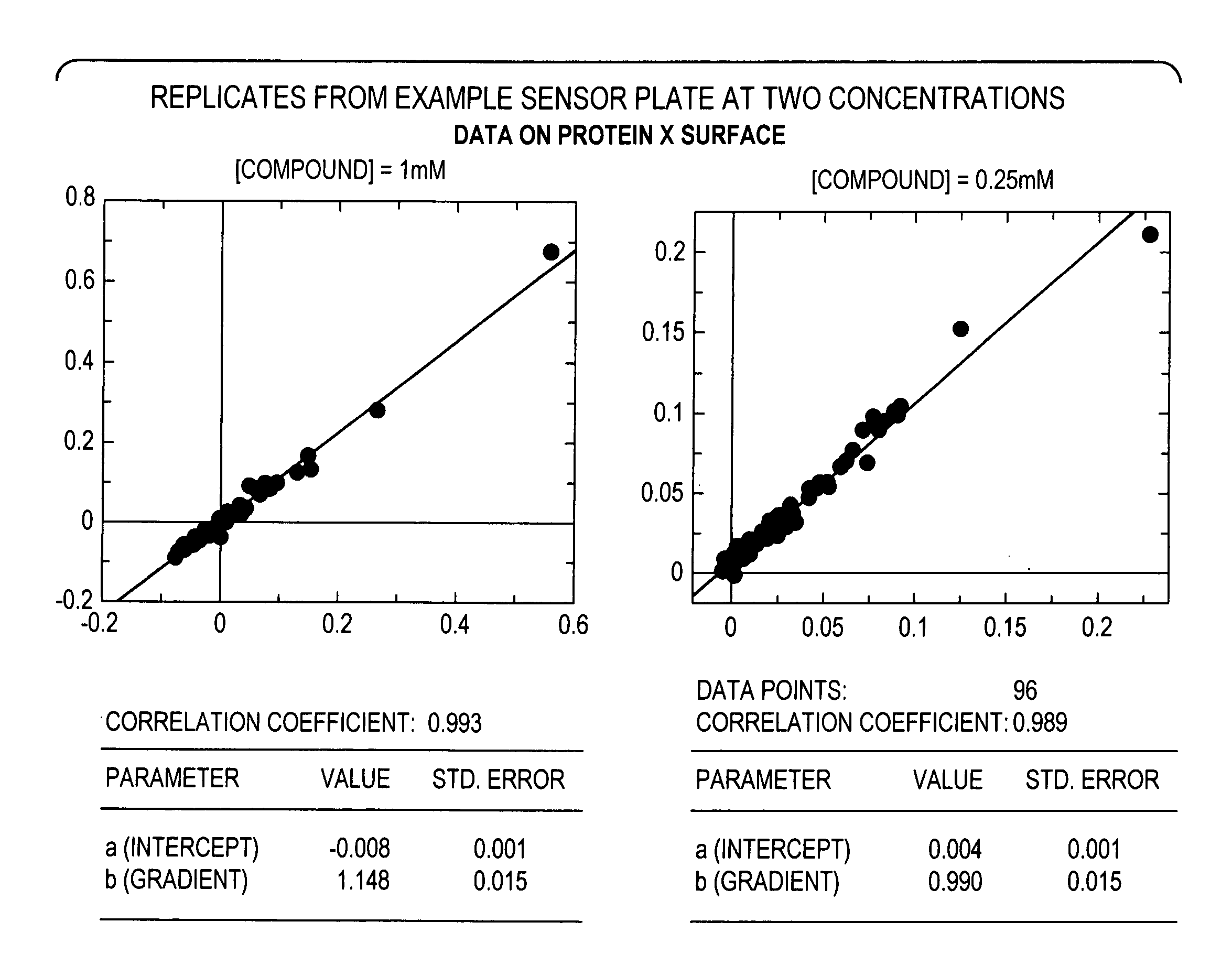
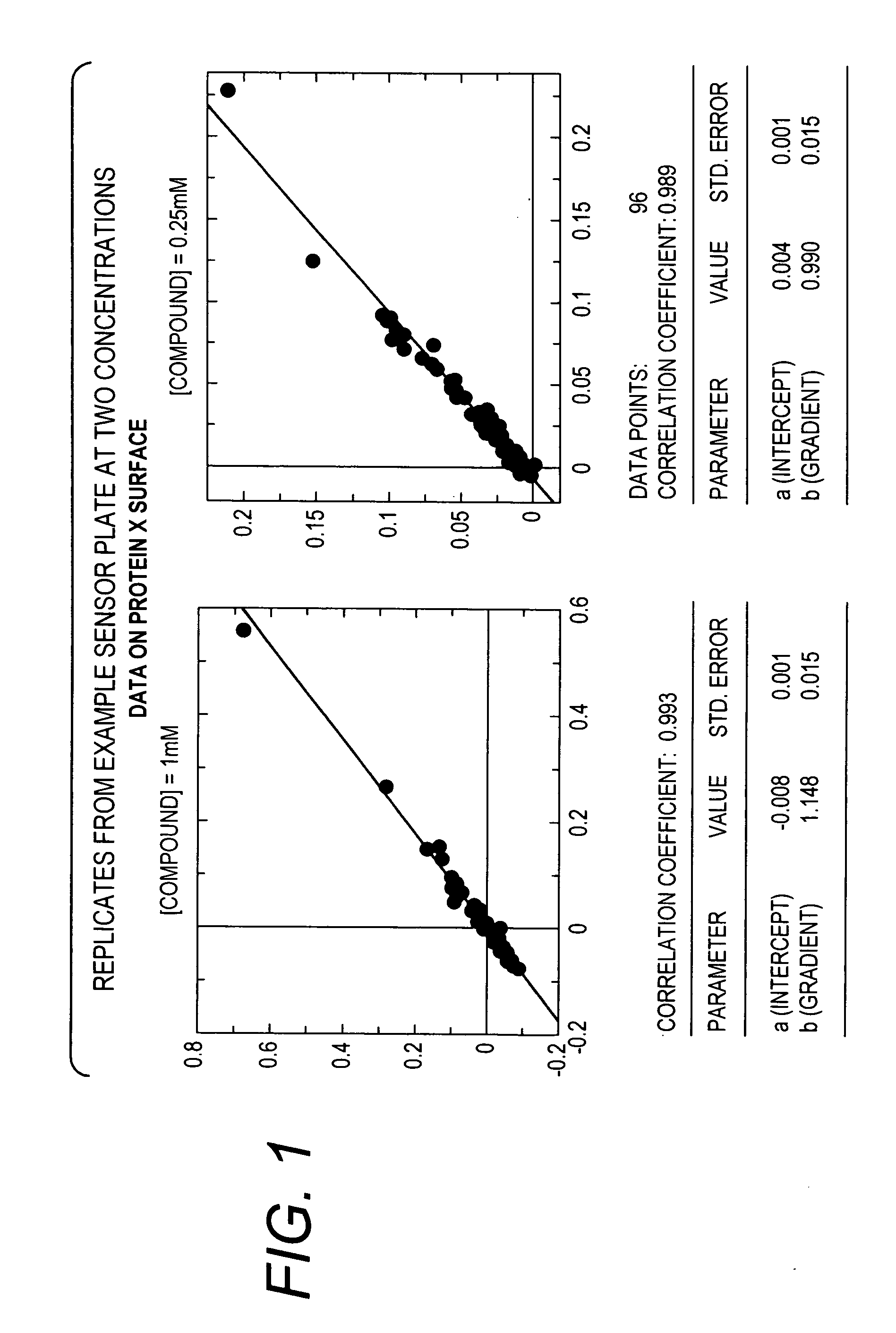
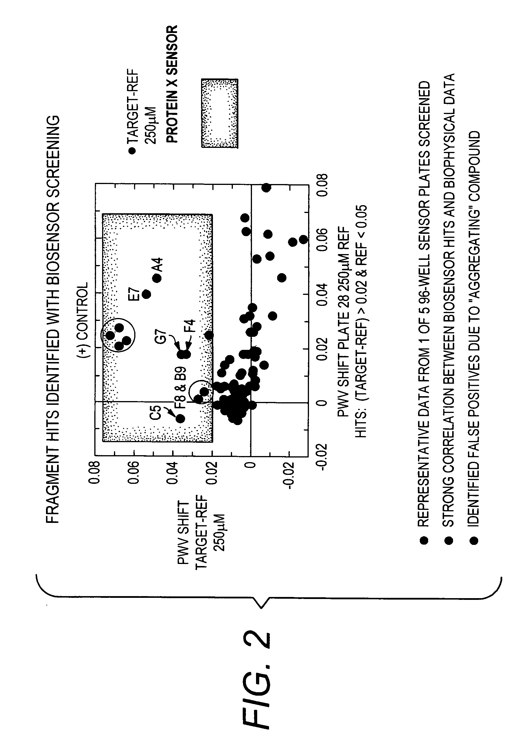
[0064]A target molecule, Protein X (26 kDa), having an ATP-ase domain, was immobilized to a colorimetric resonant reflectance biosensor. A compound library having an average MW of library members of <300 Da was added to the biosensor. Direct binding of the library constituents to the biosensor was detected. Binding data for the compound library was also obtained from biophysical methods, including NMR and X-ray crystallography.

[0065]About 500 molecules in 2% DMSO buffer were screened. The read time was less than 10 minutes. The ligand screen concentrations were 250 μM and 1 mM. The results showed: a dynamic range for 200 Da ligand with 1:1 stoichiometry, full binding 60 pm, S / N=12; proper identification of positive and negative controls; a range of affinities detected / titrated 1 nM to ˜1 mM; and strong hit correlation with lower throughput biophysical methods.

[0066]The following compounds were identified (see FIGS. 2 and 3):

[0067]Fragment A: (Mr=265 Da, Kd˜0.01 uM)

[0068]Fragment B: ...


PUM


Abstract

The invention provides method of detecting interactions of small molecules with target molecules.

Description

PRIORITY[0001]This application claims the benefit of U.S. Pat. No. 60 / 912,725, filed on Apr. 19, 2007, which is incorporated herein by reference in its entirety.BACKGROUND OF THE INVENTION[0002]Identification of high quality lead compounds for druggable protein targets is a complex task. It can be difficult to generate leads that are novel and optimizable. Recently, fragment-based screening has been explored as a method of generating high quality leads. It appears that compounds with a molecular weight of about 100 Da to about 300 Da (i.e., “fragments”) may provide better leads than compounds with a molecular weight of about 300 Da to about 600 Da, which have historically been used for lead generation. Although fragments are often weakly binding molecules, screening for weakly binding molecules of low molecular weight that bind to therapeutic target proteins is useful to determine small building blocks that can be chemically combined to provide tighter binding compounds of slightly ...

Claims


Application Information

Patent Timeline
IPC IPC(8): G01N21/62
CPCG01N21/7743G01N21/7746G01N33/54373G01N2021/7773
Inventor WAGNER, RICKLAING, LANCE
Owner X BODY
Learn More