In vivo optical imaging

a technology of optical imaging and in vivo, which is applied in the field of fluorescence, can solve the problems of limited penetration of light through the body, and achieve the effect of facilitating cleavag

Inactive Publication Date: 2009-01-01
GENERAL ELECTRIC CO
View PDF3 Cites 12 Cited by
  • Summary
  • Abstract
  • Description
  • Claims
  • Application Information

AI Technical Summary

Problems solved by technology

The major limitation of light emitting probe is the high absorption and scattering that occur in biological tissues, which cause limited penetration of the light through the body.

Method used


Image

  • In vivo optical imaging
  • In vivo optical imaging
  • In vivo optical imaging

Examples


embodiments

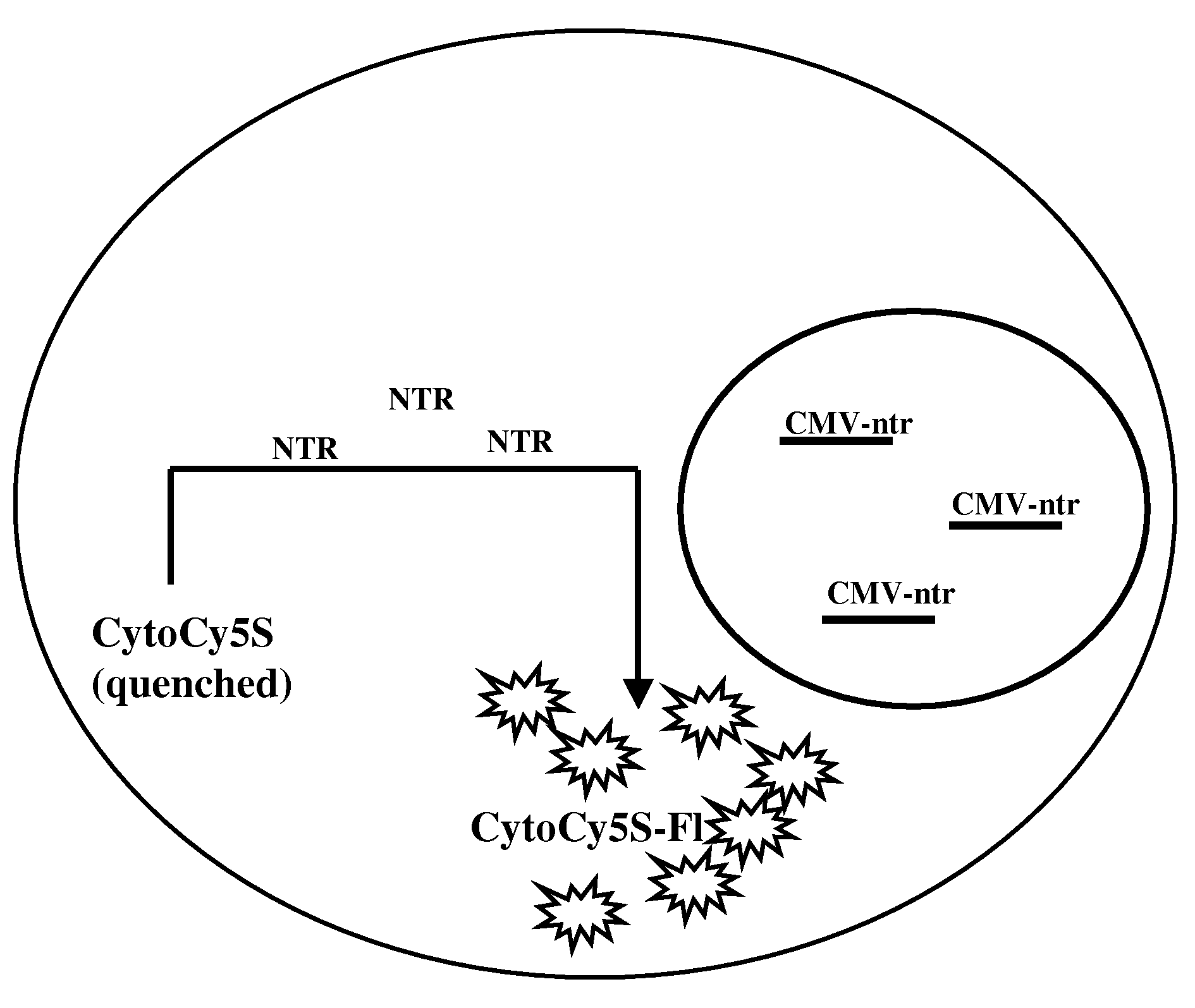
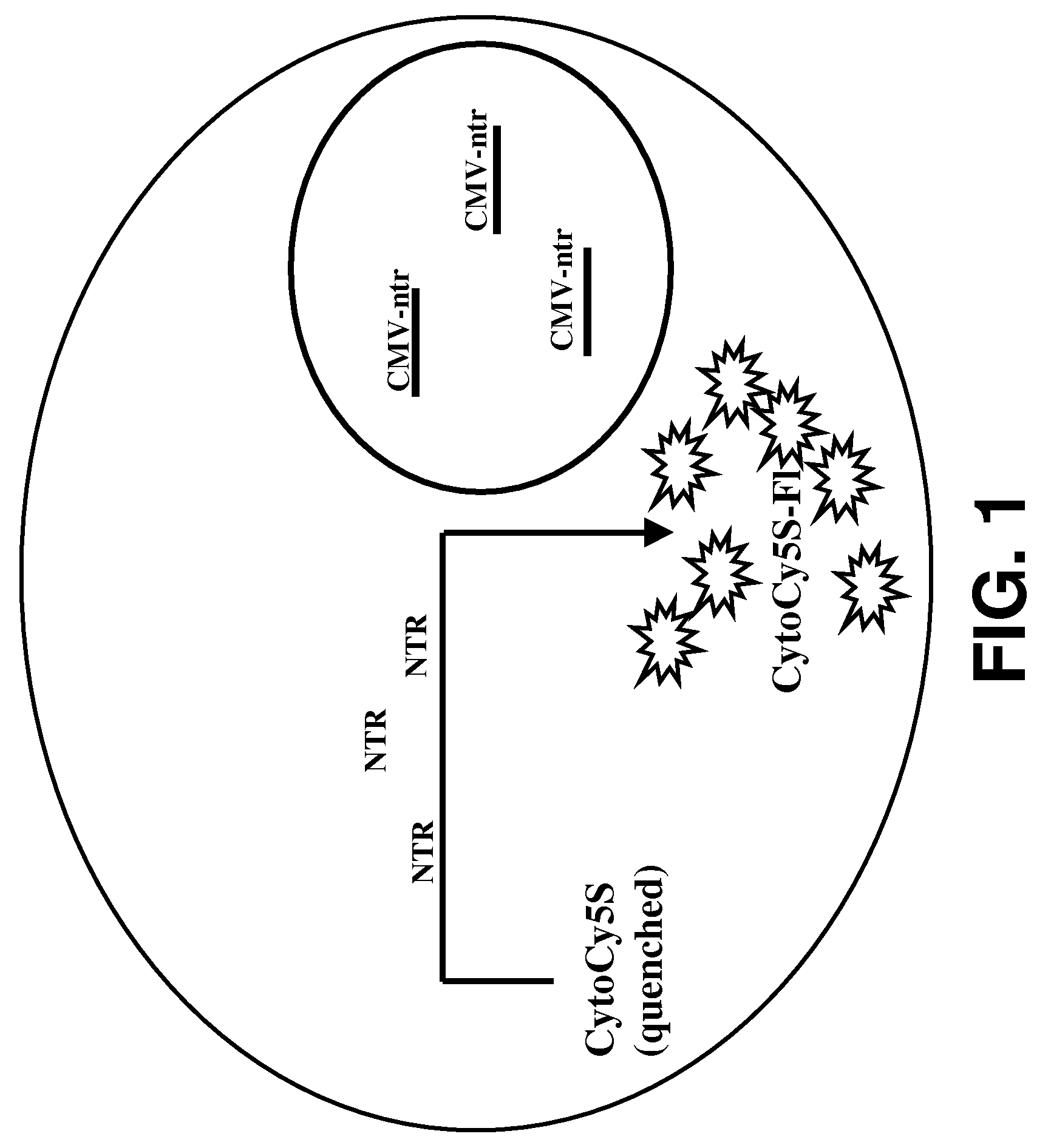
[0029]Provided herein are methods for imaging an optically labeled target cell implanted in vivo using a reporter construct that encodes an enzyme that is not endogenous to the target cell. The disclosed methods include the steps of: (a) introducing target cell transformed with non-endogenous enzyme into a mammalian subject; (b) contacting the transformed target cell with a cell-permeable soluble fluorescent dye; and (c) observing a fluorescent signal generated by the target cell present in the mammalian subject. In some embodiments, the non-endogenous enzyme comprises a nitroreductase (e.g., NTR). In some embodiments, the methods may also include the step of tracking the fluorescent target cells in the mammalian subject over time.

Nitroreductase Gene Expression

[0030]For use as a reporter gene, the nitroreductase gene may be isolated using art-recognized methods, for example by amplification from a cDNA library by use of the polymerase chain reaction (PCR). Once isolated, the nitrore...

example 1

Transformation of the Target Cells with the NTR Gene

[0075]Human colorectal carcinoma cell (LS174T cells), 2×10⁶ were plated in 60 mm Petri plate and allowed to attain 70% confluency. The cells were transfected with CMV-ntr using Superfect reagent (Qiagen, Valencia, Calif.) following the protocol of stable transfection recommended by the manufacturer. The transfected cells were selected and maintained under selective media. The stable cell line was named as LS174T-NTR. In vitro live cell assay, using CytoCy5S revealed that the cells were stably transfected by NTR reporter gene. LS174T transfected with NTR genes shows fluorescent signal from cells when incubated with CytoCy5S.

example 2

Substrate Selection

[0076]CytoCy5S dye (GE Healthcare) in powder form (1 mg) was dissolved in DMSO (100 μl) and further diluted in cell culture media 1:100 before adding to 2 cell lines SKOV control (SKOV-NTR−) and SKOV-NTR stably transformed (SKOV-NTR+) cells and LS174T control (LS174T-NTR−) and LS174T-NTR stably transformed (LS174T-NTR+). Both cell types were cultured in 96-well plate with 0.5 ng / well of CytoCy5S dye. Fluorescent signal emitted from the cells was confirmed using a Nikon TE2000-U fluorescent microscope and Incell 1000.


PUM


Abstract

Provided herein are methods for imaging an optically labeled target cell implanted in vivo using a reporter construct that encodes an enzyme that is not endogenous to the target cell. The disclosed methods include the steps of: (a) introducing target cell transformed with non-endogenous enzyme into a mammalian subject; (b) contacting the transformed target cell with a cell-permeable soluble fluorescent dye; and (c) observing a fluorescent signal generated by the target cell present in the mammalian subject. In some embodiments, the non-endogenous enzyme comprises a nitroreductase. In some embodiments, the methods may also include the step of tracking the fluorescent target cells in the mammalian subject over time.

Description

BACKGROUND[0001]The use of fluorescence as a detection modality in biological assays is widespread and a diverse variety of procedures are available to generate fluorescence under assay conditions for detection by techniques such as fluorescence microscopy, fluorescence immunoassay and flow cytometry. Fluorescent signals may be generated using an enzyme to convert a non-fluorescent or substantially non-fluorescent substrate into a fluorescent product.[0002]Such fluorescent enzyme substrates typically have two components that are coupled through a covalent linkage. One component is a fluorescent molecule that is capable of fluorescing by first accepting light energy and then emitting light energy. The other component is a masking group that prevents the fluorescent molecule from accepting or emitting light energy when the two components are covalently bound to one another, such that the molecule is non-fluorescent or substantially non-fluorescent. In the presence of an appropriate en...

Claims


Application Information

Patent Timeline
Patent Type & Authority Applications(United States)
IPC IPC(8): A61K49/00A61P43/00
CPCA61K49/0032A61P43/00
Inventor BHAUMIK, SRABANIAGDEPPA, ERIC DUSTINKLIMASH, JUNEDEPUY, JEANNETTE CHRISTINE
Owner GENERAL ELECTRIC CO
Why Eureka
Try Eureka