Method for determination of bone fracture risk using raman spectroscopy

a raman spectroscopy and risk technology, applied in the field of detecting the risk of bone fracture, can solve the problems of abnormal porous bone, poor prediction of osteoporosis, and often normal bone density of sufferers of osteoporosis fractures, and achieve the effect of rapid determination of the risk of osteoporosis and non-invasiveness

Inactive Publication Date: 2009-01-08
UNIVERSITY OF WYOMING +1
View PDF1 Cites 9 Cited by
  • Summary
  • Abstract
  • Description
  • Claims
  • Application Information

AI Technical Summary

Benefits of technology

[0012]The present invention can be used as a diagnostic aid for assessing the risk of disease such as osteoporosis and the corresponding bone fracture risk. The Raman imaging technique of the invention provides a non-invasive and rapid determination of the risk of osteoporosis and bone fracture risk in live subjects. The present invention thus offers valuable diagnostic information applicable to large populations that may help in assessing an individual's risk of having bone fracture, and in determining protocols for prevention of the same. In a preferred embodiment of the present invention, the method is automated and carried out in a single instrument without the need for an operator. Accordingly, a lay subject wishing to be tested for loss of bone strength can place the keratinized tissue sample, either removed from the subject or still resident on the subject, into the instrument and the instrument will automatically carry out the method and provide an output to the subject indicative of the bone health of the subject. Alternatively the instrument may be used by a health care practitioner to analyze samples of keratinized tissue of a subject under the care or treatment of the health care professional.

Problems solved by technology

Osteoporosis is a condition that features loss of the normal density of bone and leads to abnormally porous bone.
Osteoporosis is however a poor predictor of bone fracture the clinical endpoint.
Unfortunately, patients who suffer osteoporotic fractures often have normal bone density, because it is the organic phase, as well as the mineral phase, that is affected.
Bone densitometry is an involved and expensive method to measure bone density.
Central devices are more accurate, but cost significantly more than peripheral devices.
It is now, however, fairly well accepted that bone fracture risk can derive from protein problems.
Bone fragility may result from a disturbance, to variable degree, of these collagenous and non-collagenous bone proteins.
The problem with Limerick's approach is that it is very difficult to measure the width (more specifically the “full width at half maximum) of a peak.
This can be difficult to measure since natural materials such as fingernails have significant fluorescent backgrounds.

Method used


Image

  • Method for determination of bone fracture risk using raman spectroscopy
  • Method for determination of bone fracture risk using raman spectroscopy
  • Method for determination of bone fracture risk using raman spectroscopy

Examples


Embodiment Construction

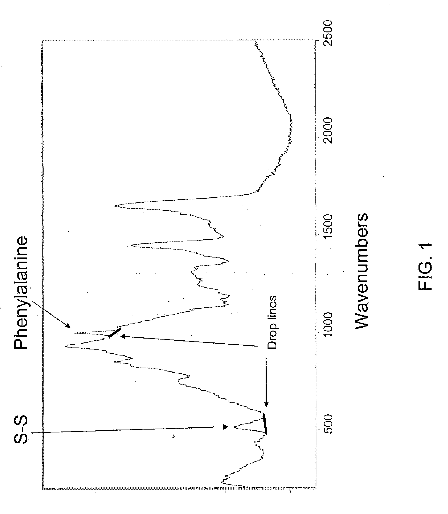
[0021]Raman spectroscopy is the measurement of the wavelength and intensity of inelastically scattered light from molecules. The Raman scattered light occurs at wavelengths that are shifted from the incident light by the energies of molecular vibrations. The mechanism of Raman scattering is different from that of infrared (IR) absorption, and Raman and IR spectra provide complementary information. Raman spectroscopy is commonly used in chemistry, since vibrational information is very specific for the chemical bonds in molecules. It therefore provides a fingerprint by which the molecule can be identified. Typical applications are in structure determination, multi-component qualitative analysis, and quantitative analysis. An apparatus for performing Raman spectroscopy in the detection of carotenoids in the macular tissue of a subject is described in U.S. Pat. No. 7,039,452, which is incorporated herein by this reference. An apparatus for performing Raman spectroscopy in the detection ...


PUM

PropertyMeasurementUnit
fracture riskaaaaaaaaaa
peak heightaaaaaaaaaa
heightaaaaaaaaaa

Abstract

A method is disclosed for analyzing keratinized tissue, particularly fingernails, of a subject to diagnose osteoporosis and bone fracture risk. A Raman spectrum of a sample of keratinized tissue is generated. Broad spectral background features of the spectrum are removed, preferably by using Fourier transform analysis. Peak heights of Raman features of interest, particularly the S—S bond of cystine, are measured. These peak height measurements are normalized using reference peak heights of Raman features that are invariant between normal and osteoporotic subjects, such as the CH2 bending peak.

Description

BACKGROUND OF THE INVENTION[0001]The invention relates generally to methods for detecting the risk of bone fracture and, more specifically, to a method for determining the existence of low bone quality using Raman spectroscopy.[0002]Osteoporosis is a condition that features loss of the normal density of bone and leads to abnormally porous bone. Osteoporosis affects over 44 million Americans and over one-half of these are women. The exact cause of osteoporosis is unknown. Since bones are composed of a mixture of an organic phase, primarily collagen, and an inorganic phase, primarily calcium phosphate, a prevailing thought has been that calcium deficiency leads to osteoporosis. The propensity of osteoporosis in women supports this conclusion since they often lose calcium from lactation and throughout menopause. Osteoporosis is however a poor predictor of bone fracture the clinical endpoint. The need for Bone Quality based assessments that may have improved prediction capability is wel...

Claims


Application Information

Patent Timeline
Patent Type & Authority Applications(United States)
IPC IPC(8): A61B6/00G01N21/65
CPCA61B5/0059A61B5/4504G01N21/65A61B5/7257A61B5/7275A61B5/4509G16H30/40G16H50/20G16H50/30
Inventor CARRON, KEITHTOWLER, MARK
Owner UNIVERSITY OF WYOMING
Why Eureka
Try Eureka