

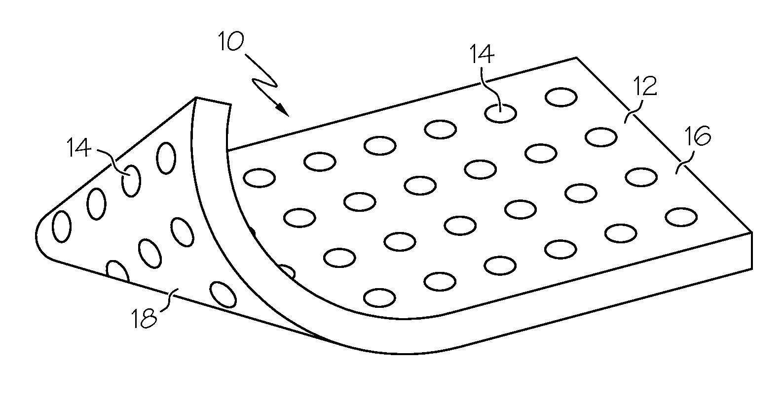
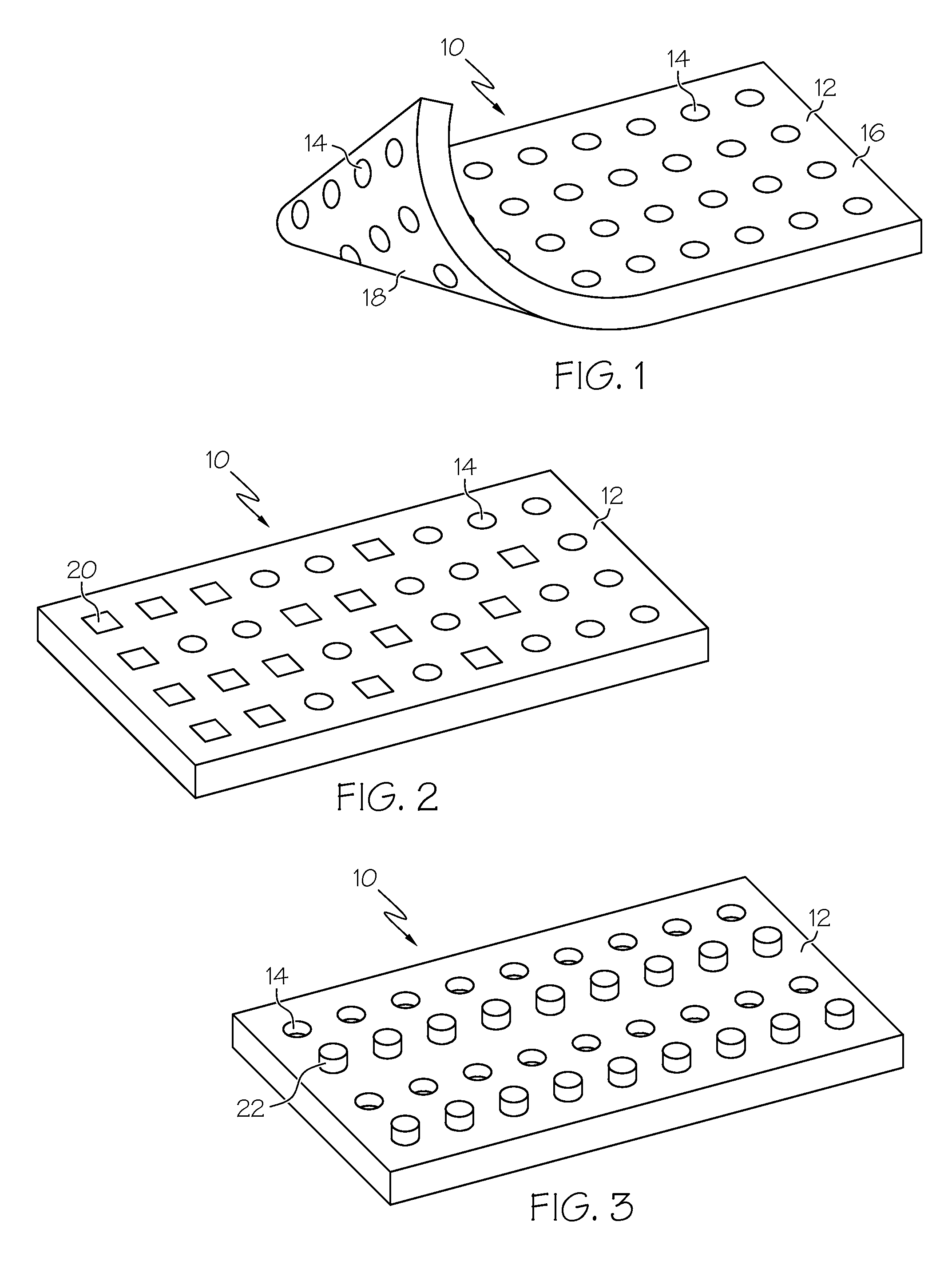
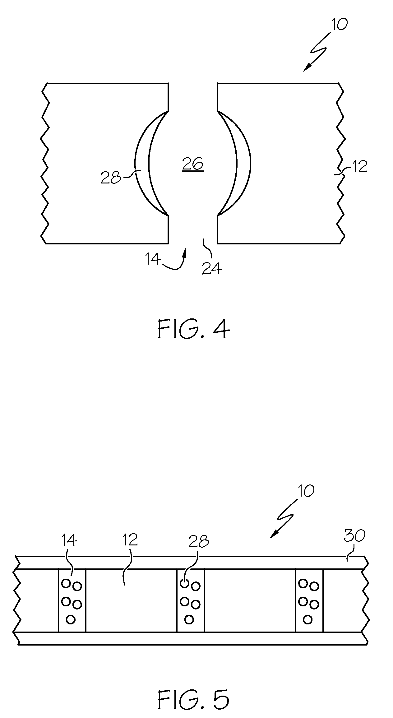
Biomaterial including micropores
a technology of biomaterials and micropores, applied in the field of biomaterials including micropores, can solve the problems of foaming altering the composition of biomaterials used, unavailability, and undesirable physiological effects
- Summary
- Abstract
- Description
- Claims
- Application Information
AI Technical Summary
Benefits of technology
Problems solved by technology
Method used
Image

Examples
example 1
[0065]A series of experiments were conducted over a four-week time period in the dog animal model. The dog had implanted silicone biomaterial sheet with 500 micron pores with a 0.020 inch thickness that was laid onto the body dorsum of the dog's nose. The histological results indicated that there was complete tissue in-growth into the pores and incorporation of soft tissue as well as excellent vascularization with only a limited inflammatory response. Vasculature in some cases permeated the entire thickness of the membrane through the micropores extending from one surface to the other. In contrast, a control material without pores had classic fiber capsule formation around it with perivasculature outside the capsule and no integration of the implant with the surrounding tissue. There was no evidence of seroma formation, infection, or extrusion.
example 2
[0066]A series of experiments were also conducted over a four-week time period in the rabbit animal model. In the case of the rabbit animal model, a segment of ear cartilage was removed and replaced with two sheets of Silastic® biomaterial that had 250 micron pores through the sheeting. This was implanted subcutaneously as a cartilaginous replacement. In the rabbit experiment, the histologic results indicated no seroma formation had occurred post operatively with intimate integration of the soft tissue into and through the pores along with excellent vascularity throughout all the pores of the implant. Where some areas of the implant overlapped the rabbit's ear cartilage, the material displayed good tissue integration with no evidence of cartilaginous necroses or disruption of the cartilage microstructure. There was no evidence of seroma formation, infection, or extrusion.
PUM
| Property | Measurement | Unit |
|---|---|---|
| Diameter | aaaaa | aaaaa |
| Diameter | aaaaa | aaaaa |
| Diameter | aaaaa | aaaaa |
Abstract
Description
Claims
Application Information