Quantitative clinical and pre-clinical imaging

a quantitative, clinical technology, applied in the field of medical science, can solve the problems of heightened problems, inability to accurately predict the effect of clinical outcomes, so as to improve the efficiency of design, improve the value of clinical outcomes, and improve the efficiency of image registration

Inactive Publication Date: 2009-04-02
KONINKLIJKE PHILIPS ELECTRONICS NV
View PDF15 Cites 19 Cited by
  • Summary
  • Abstract
  • Description
  • Claims
  • Application Information

AI Technical Summary

Benefits of technology

[0015]One advantage resides in enhanced value in clinical and preclinical studies due to automated generation of error and confidence interval information.
[0017]Another advantage resides in improved efficiency in the design and implementation of clinical and preclinical studies.
[0018]Another advantage resides in the combination of in-vivo imaging data with in-vitro measurements, in-silico results, and / or ex-vivo histology to assess and / or improve statistical significant results.

Problems solved by technology

Error, in the form of noise, lack of precision, and / or uncontrolled variation of whatever kind can be introduced at substantially any stage of the processing including during imaging data acquisition, image reconstruction, post-reconstruction image processing, multimodality spatial image registration, extraction of clinically significant results from reconstructed images, and so forth.
In practice, however, these estimates are typically performed manually using office-type spreadsheets such as Microsoft® Excel or other manually operated calculation aids, and in some such cases calculation of the resulting statistical significance with each form of error taken into account is either not done or itself subject to error.
Failure to determine, record, and preserve such information relevant to error or confidence interval assessment can lead to expensive and time-consuming pre-clinical or clinical studies that are fundamentally flawed and of limited or non-existent value for making informed decisions regarding the experimental therapy or diagnostic under investigation.
These problems are heightened when the study employs multimodality imaging.
However, it is difficult or impossible using existing techniques to empirically determine the amount of error introduced by these registration operations.
Existing calibration phantoms have substantial deficiencies and are not effective for calibrating, or assessing error introduced by, image registration techniques.
In view of the increasingly common use of multimodality imaging in pre-clinical and clinical studies, this fundamental deficiency in image registration is problematic because it introduces error of largely unknown magnitude, nature, and effect on error propagation into the study analyses.

Method used


Image

  • Quantitative clinical and pre-clinical imaging
  • Quantitative clinical and pre-clinical imaging
  • Quantitative clinical and pre-clinical imaging

Examples


Embodiment Construction

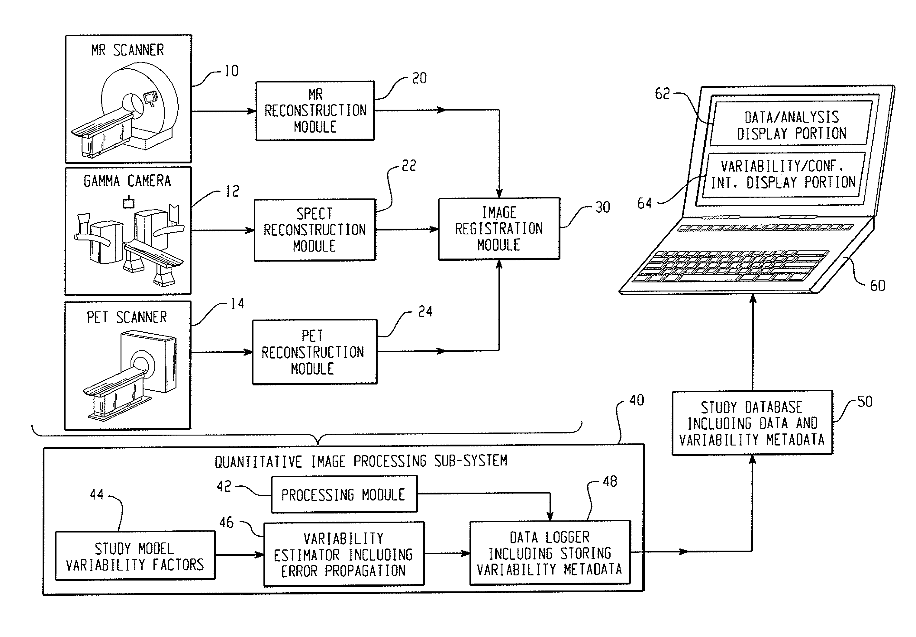
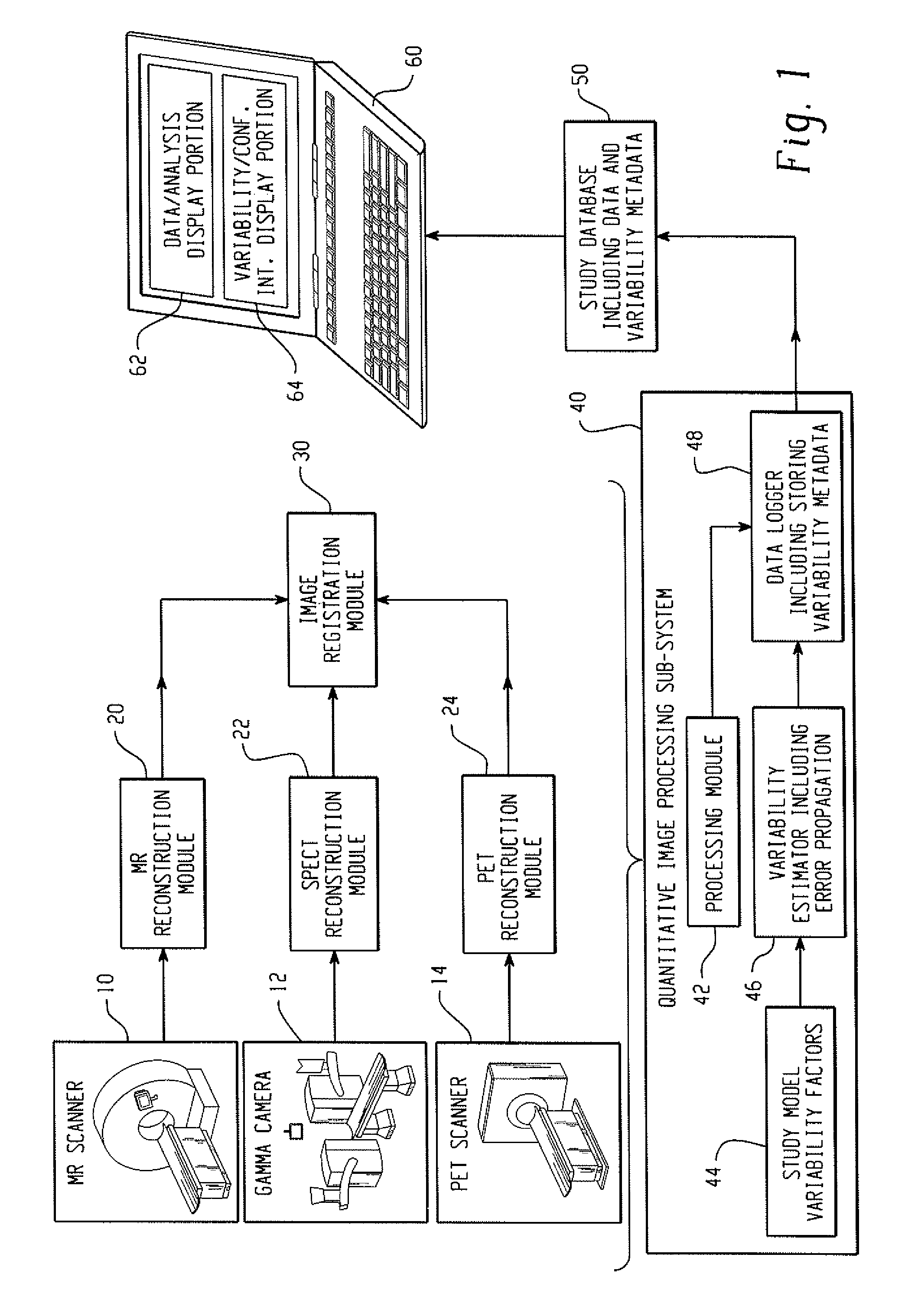
[0024]With reference to FIG. 1, a clinical or preclinical imaging system includes an image acquisition subsystem including a data acquisition element and an image reconstruction element cooperating to generate clinical or preclinical images of clinical or preclinical subjects. In illustrative FIG. 1, the clinical or preclinical imaging system includes three data acquisition elements, namely a magnetic resonance (MR) scanner 10, a gamma camera 12 for single photon emission computed tomography (SPECT) data acquisition, and a positron emission tomography (PET) scanner 14. The illustrative clinical or preclinical imaging system also includes three corresponding image reconstruction elements, namely an MR reconstruction module 20, a SPECT reconstruction module 22, and a PET reconstruction module 24. These illustrative components are examples; more generally, the clinical or preclinical imaging system can include as few as a single data acquisition element and corresponding reconstruction...


PUM


Abstract

In a clinical or preclinical imaging system, an image acquisition subsystem includes a data acquisition and image reconstruction elements generating clinical or preclinical images. A quantitative image processing subsystem generates variability metadata associated with the clinical or preclinical images, and a clinically or preclinically significant result with an associated confidence interval computed based on the variability metadata. A user interface displays the clinically or preclinically significant result together with the associated confidence interval. A phantom for calibrating such an imaging system includes a deformable nonbiological structure approximating structure of a clinical or preclinical subject to be imaged, and fiducial markers detectable by the imaging system disposed on or in the deformable nonbiological structure to move with deformation of the deformable nonbiological structure.

Description

CROSS REFERENCE TO RELATED APPLICATIONS[0001]This application claims the benefit of U.S. provisional application Ser. No. 60 / 976,519 filed Oct. 1, 2007, which is incorporated by reference.BACKGROUND OF THE INVENTION[0002]The following relates to the medical arts. The following finds illustrative application to clinical and pre-clinical imaging, and is described with particular reference thereto. However, the following will find application in other medical applications such as, but not limited to, measurement of diagnostically relevant parameters to aid in patient triage and management.[0003]Medical therapies and diagnostic methods or systems in the research, development, and certification stages are typically tested pre-clinically on animals such as mice, guinea pigs, or so forth. If the pre-clinical tests are promising and indicate an acceptable level of safety, the development proceeds to clinical studies on human volunteers. Based on the results of such clinical studies, the eff...

Claims


Application Information

Patent Timeline
Patent Type & Authority Applications(United States)
IPC IPC(8): A61B5/05G06G7/60G16H30/40
CPCA61B6/5247A61B6/563A61B6/583A61B6/508G06F19/321A61B2503/40G01T1/1603G16H30/40
Inventor ZAGORCHEV, LYUBOMIRSTANTON, DOUGLASBUCKLER, ANDREWMALLYA, YOGISH
Owner KONINKLIJKE PHILIPS ELECTRONICS NV
Why Eureka
Try Eureka