Pre-clinical method for monitoring serial changes in circulating breast cancer cells in mice

a breast cancer cell and mice technology, applied in the field of cancer monitoring and assessing disease progression in metastatic cancer patients, can solve the problems of limited research on the role of ctc in metastasis and the expansion of their use as a biomarker in pharmacokinetic and pharmacodynamic studies, and is difficult to detect and elimina

Inactive Publication Date: 2009-05-07
VERIDEX LCC
View PDF10 Cites 5 Cited by
  • Summary
  • Abstract
  • Description
  • Claims
  • Application Information

AI Technical Summary

Problems solved by technology

Research on the role of CTC in metastasis and expansion of their use as a biomarker in pharmacokinetic and pharmacodynamic studies has been limited to the clinical phase of drug development.
Unfortunately, the metastatic colonies are difficult to detect and eliminate and it is often impossible to treat all of them successfully.
Assessing enzyme activity in this type of analysis can involve time-consuming laboratory procedures such as gel electrophoresis and Western blot analysis.
However in both cases, the base levels during remission, or even in healthy normals, are relatively high and may overlap with concentrations found in patients, thus requiring multiple testing and monitoring to establish patient-dependent baseline and cut-off levels.
However, PSA or the related PSMA testing leaves much to be desired.
For example, elevated levels of PSA weakly correlate with disease stage and appear not to be a reliable indicator of the metastatic potential of the tumor.
This is being done through the laborious procedure of isolating all of the mRNA from the blood sample and performing reverse transcriptase PCR.
Additionally, false positives are often observed using this technique.
There is an added drawback, which is that there is a finite and practical limit to the sensitivity of this technique based on the sample size.
The aforementioned studies, while seemingly prognostic under the experimental conditions, do not provide for consistent data with a long follow-up period or at a satisfactory specificity.
Accordingly, these efforts have proven to be somewhat futile as the appearance of mRNA for antigens in blood have not been generally predictive for most cancers and are often detected when there is little hope for the patient.
These results suggest that tumor cells were shed into the bloodstream (possibly during surgical procedures or from micro metastases already existing at the time of the operation), and resulted in poor patient outcomes in patients with colorectal cancer.
As mentioned, these detection ranges are based on unreliable conversions of amplified product to the number of tumor cells.
Further, PCR-based assays are limited by possible sample contamination, along with an inability to quantify tumor cells.
Most importantly, methods based on PCR, flowcytometry, cytoplasmic enzymes and circulating tumor antigens cannot provide essential morphological information confirming the structural integrity underlying metastatic potential of the presumed CTC and thus constitute functionally less reliable surrogate assays than the highly sensitive imaging methods embodied, in part, in this invention.
Unfortunately, the same spreading of malignant cells continues to be missed by conventional tumor staging procedures.
But these invasive techniques are deemed undesirable or unacceptable for routine or multiple clinical assays compared to detection of disseminated epithelial tumor cells in blood.
Currently available preclinical protocols have not demonstrated a consistently reliable means for repetitively monitoring CTC's in assessing metastatic breast cancer (MBC) progression.
The inability to repetitively monitor CTC's in the small blood volumes available in pre-clinical animal models of breast and other cancers has restricted their use to analysis of samples obtained from terminal blood draws.

Method used


Image

  • Pre-clinical method for monitoring serial changes in circulating breast cancer cells in mice
  • Pre-clinical method for monitoring serial changes in circulating breast cancer cells in mice
  • Pre-clinical method for monitoring serial changes in circulating breast cancer cells in mice

Examples


example 1

Enumeration of Circulating Cytokeratin Positive Cells

[0032]The CellTracks® System refers to an automated fluorescence microscopic system for automated enumeration of isolated cells from blood. The system contains an integrated computer controlled fluorescence microscope and automated stage with a magnetic yoke assembly that will hold a disposable sample cartridge. The magnetic yoke is designed to enable ferrofluid-labeled candidate tumor cells within the sample chamber to be magnetically localized to the upper viewing surface of the sample cartridge for microscopic viewing. Software presents suspect cancer cells, labeled with antibodies to cytokeratin and having epithelial origin, to the operator for final selection.

[0033]While isolation of tumor cells for the CellTracks® System can be accomplished by any means known in the art, one embodiment uses immunomagentic enrichment for isolating tumor cells from a biological sample. Epithelial cell-specific magnetic particles are added and ...

example 2

In Vitro Recovery of Human Epithelial Cells

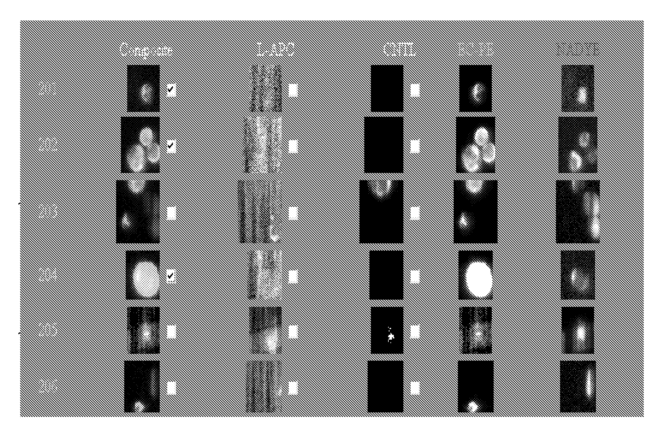
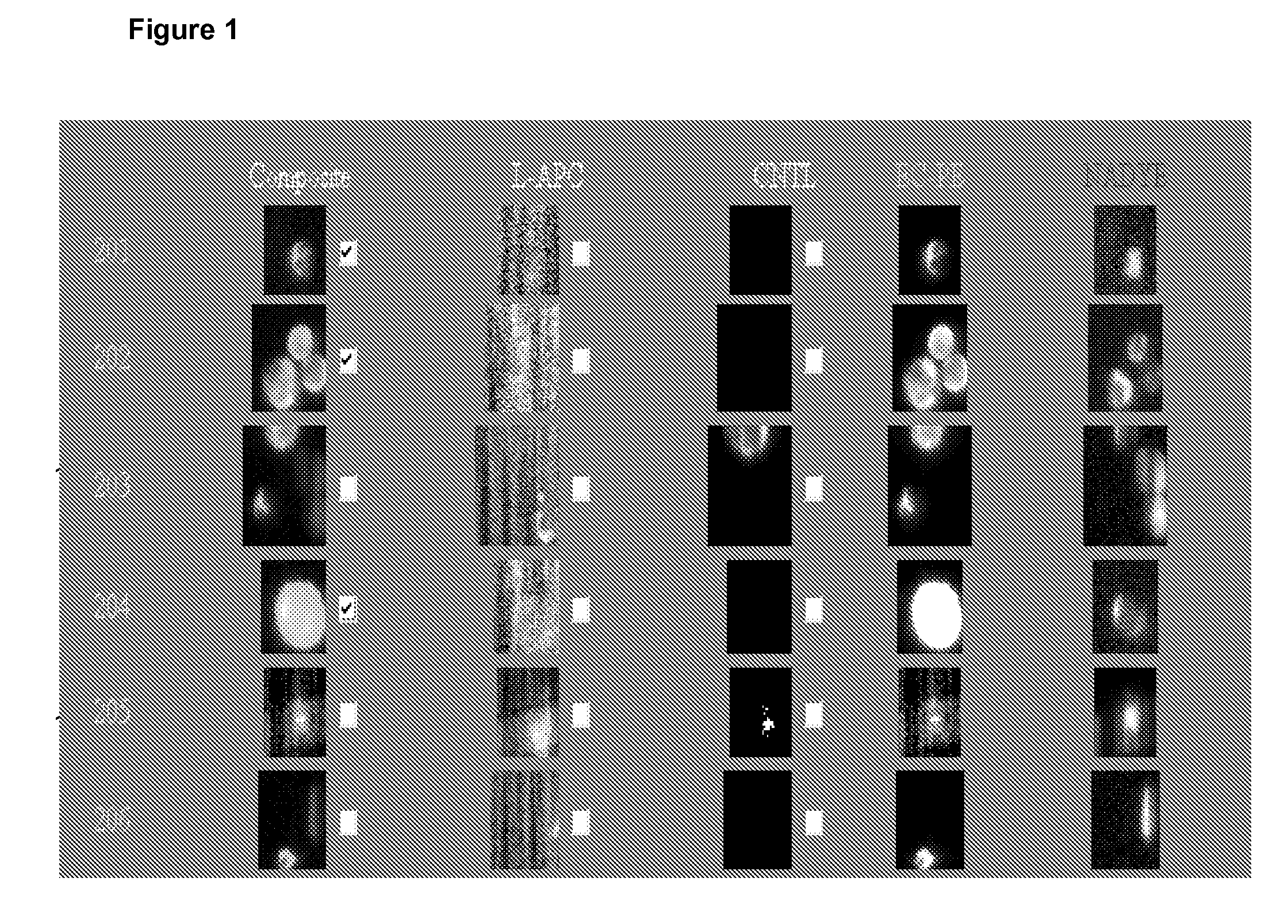
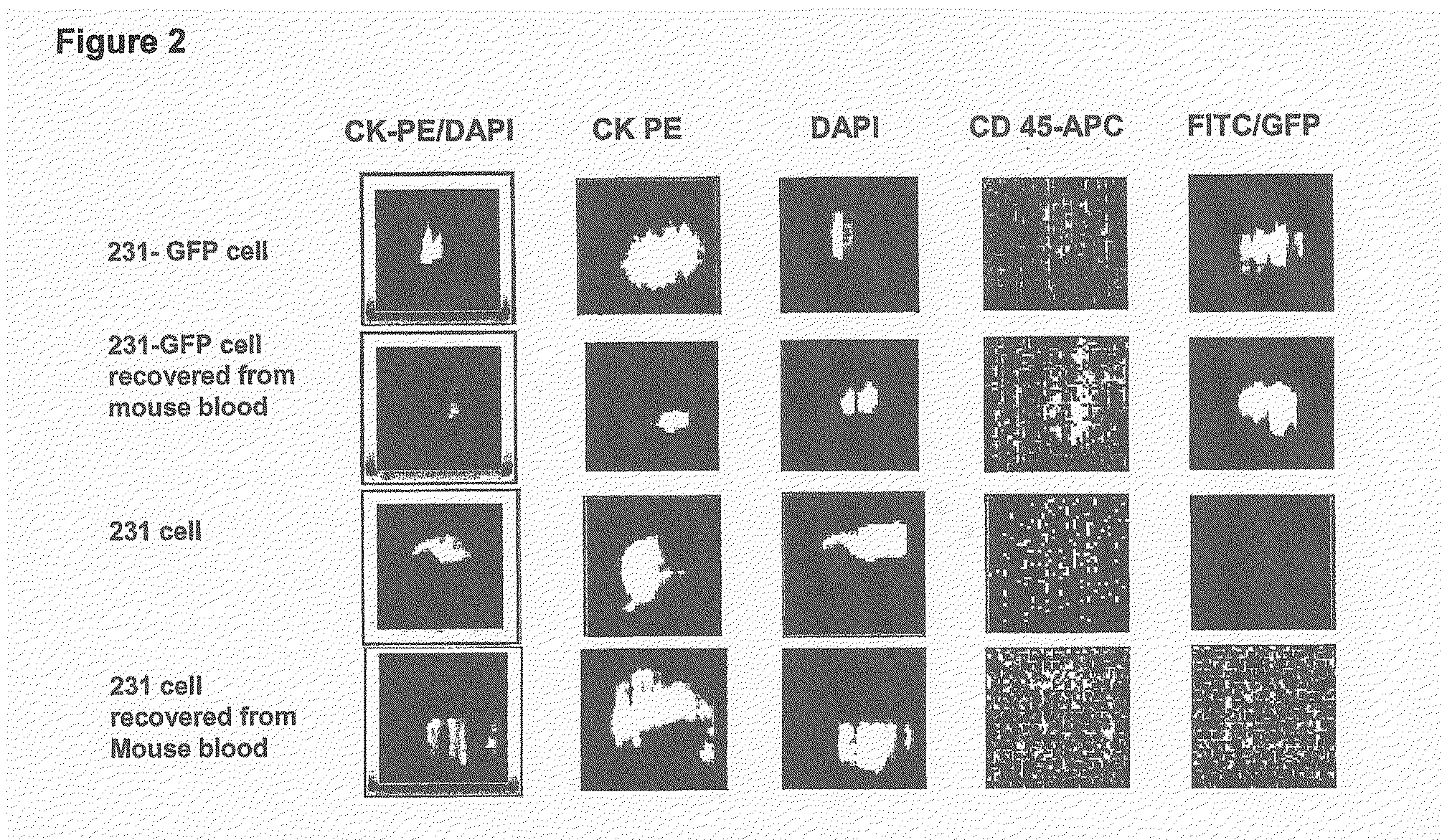
[0035]To accomplish this, 500 MDA-MB-231 breast cancer cells were spiked into 100 μl blood samples collected from mice without tumors. Since the clinical version of the assay requires blood be drawn into a proprietary vacuum tube, such as the CellSave tube, containing both an anticoagulant and a preservative, a proportionately reduced amount of CellSave solution was added to the specimens. The spiked specimens were then prepared, the CTC quantified and the percent recovery calculated. As a positive control, additional samples using MDA-MB-231 cells stably transduced with GFP were prepared. Fluorescence from GFP was detected in an open channel (FITC) of the system to confirm that all cells quantified as epithelial cells corresponded with 231-GFP cells added to mouse blood. As a negative control, mouse blood samples without cancer cells were collected, processed in an identical manner and analyzed. Of the 500 cells added to mouse blood (n=4 s...

example 3

Recovery of CTC from Xenografts in Mice

[0036]The preferred method to serially monitor CTC's in mouse models of human breast cancer incorporates the use of the CellTracks® System. As previously discussed, the system uses immunomagnetic isolation of epithelial cells from blood and immunofluorescent staining to further differentiate epithelial cancer cells from leukocytes. Because the CellTracks® system was originally developed to process 7.5 to 30 ml human blood samples, it is necessary that human epithelial breast cancer cells could be reliably recovered from small volumes of mouse blood using this assay (see Example 2).

[0037]The system was used to identify CTC's that spontaneously intravasate into the circulation from orthotopic tumor xenografts of MDA-MB-231 cells. 0.7 to 1 ml blood samples were collected from each mouse by puncture of the left ventricle when animals were euthanized for tumor burden at 10 weeks. Total numbers of CTC's ranged from approximately 100 to 1000 cells per...


PUM


Abstract

The CellTracks® System provides a system to enumerate CTC's in blood. The system immunomagnetically concentrates epithelial cells, fluorescently labels the cells and identifies and quantifies CTC's. The absolute number of CTC's detected in the peripheral blood tumor load is, in part, a factor in prediction of survival, time to progression, and response to therapy. Pre-clinical studies of circulating tumor cells (CTC's) have been limited by the inability to repetitively monitor CTC's in animal models. The present invention provides a method to enumerate CTC's in blood samples obtained from living mice, using a protocol similar to an in vitro diagnostic system for quantifying CTC's in patients. Accordingly, this technology can be adapted for serial monitoring of CTC's in mouse xenograft tumor models of human breast cancer.

Description

CROSS-REFERENCE TO RELATED APPLICATIONS[0001]This is a non-provisional application which claims priority to U.S. Provisional Applications 61 / 001,418, filed Nov. 1, 2007. The aforementioned application is incorporated in full by reference herein.BACKGROUND[0002]1. Field of the Invention[0003]The invention relates generally to cancer monitoring and assessing disease progression in metastatic cancer patients, based on the presence of morphologically intact circulating cancer cells (CTC) in blood. More specifically, methods, reagents and apparatus are described for assessing circulating cancer cells in animal models.[0004]2. Background Art[0005]Non-hematogenous epithelial tumor cells were first identified in the blood of a breast cancer patient over 150 years ago. Since then, CTC's have been shown to be a critical link between primary cancer, a disease stage at which cure is possible, and metastatic disease, which continues to be the leading cause of death for most malignancies. Clinica...

Claims


Application Information

Patent Timeline
Patent Type & Authority Applications(United States)
IPC IPC(8): A01N1/02
CPCG01N33/57415G01N33/54326
Inventor DOYLE, GERALD V.
Owner VERIDEX LCC
Why Eureka
Try Eureka