
Method and apparatus for quantifying the behavior of an administered contrast agent

a contrast agent and behavior technology, applied in the field of medical imaging, can solve the problems of difficult deconvolution analysis, unstable process, and inability to observe the tissue impulse response function r(t) directly, and achieve the effect of reducing the probability of adsorption

Active Publication Date: 2009-05-14
MULTI MAGNETICS
View PDF5 Cites 8 Cited by
  • Summary
  • Abstract
  • Description
  • Claims
  • Application Information

AI Technical Summary

Problems solved by technology

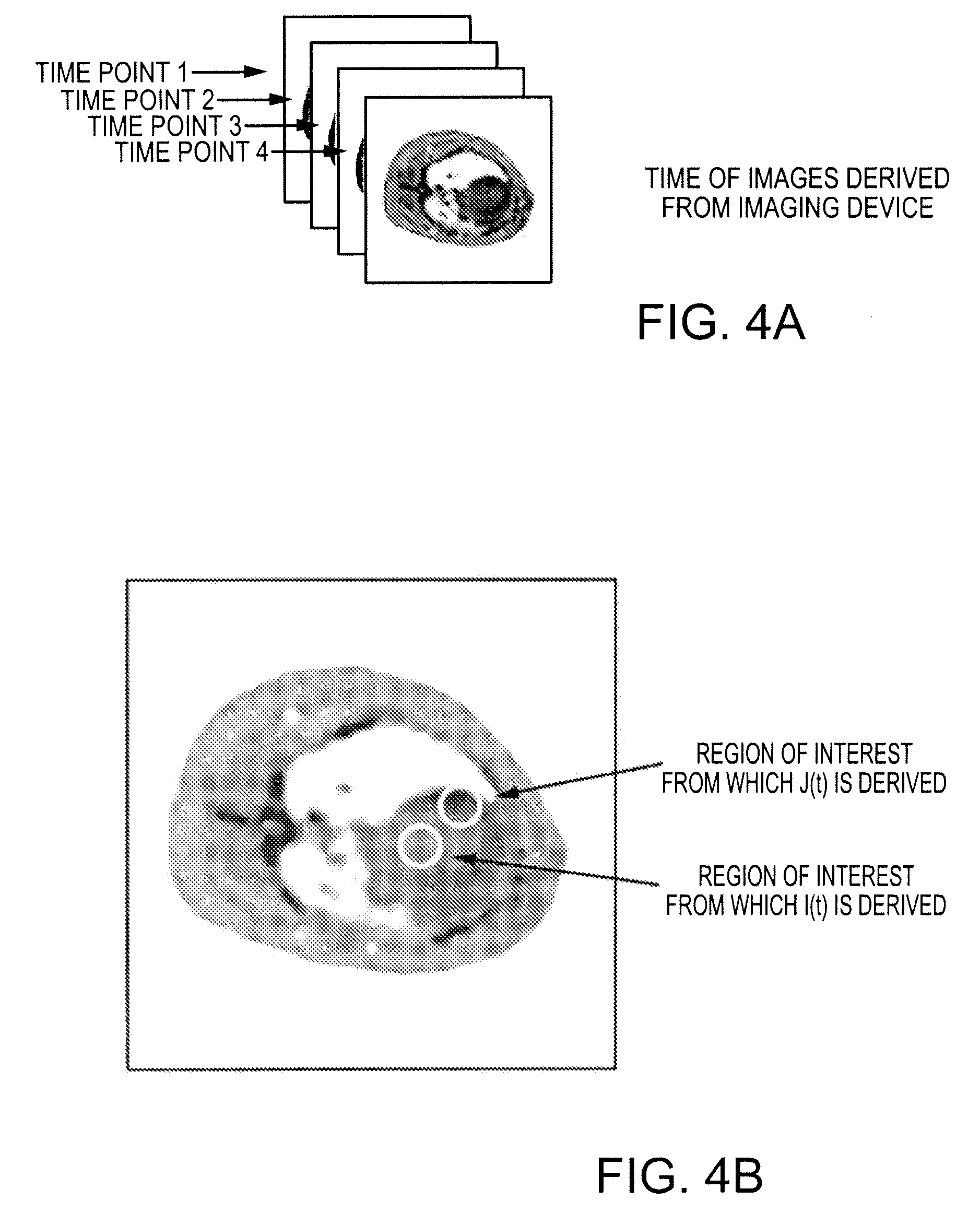
In practice, the tissue impulse response function R(t) cannot be observed directly because an ideal bolus of contrast agent cannot be directly injected into the tissue of interest.
Deconvolution analysis is difficult because it is inherently a differentiating process that is mathematically unstable and therefore very susceptible to noise in either I(t) or J(t) as described by Bronikowski et al. in “Model-free deconvolution techniques for estimating vascular transport function”, International Journal of Biomedical Engineering and Technology, Vol. 14, 411-429, 1983.
Deconvolving I(t) from J(t) in the time domain is a difficult and unstable process.
Unfortunately, significant discontinuities appear if data acquisition is terminated early because of time limitations, while a significant amount of contrast agent remains within the tissue of interest and J(t) has not yet dropped to zero as described in the aforementioned Gobbel et al. reference.
To address this concern, some authors have developed techniques to temporally extrapolate I(t) and J(t) to zero as described in the aforementioned Gobbel et al., Juni et al. and Wall et al. references; however the validity domain of temporal extrapolations of data has not been fully explored.
Reducing the number of parameters can however, limit the model as the final shape of the tissue impulse response function is restricted to the family of curves that can be realized by manipulating the parameters of the analytical model.
Analytical models work best in specific well-defined biological systems, and should not be used beyond such systems since it is difficult to know a priori the appropriate functional form as described in the aforementioned Juni et al. reference.
Unfortunately, this strategy can produce widely-oscillating R′(t) that have no physiological significance.

Method used


Image

  • Method and apparatus for quantifying the behavior of an administered contrast agent
  • Method and apparatus for quantifying the behavior of an administered contrast agent
  • Method and apparatus for quantifying the behavior of an administered contrast agent

Examples


examples

[0067]The most promising MRI technique for perfusion assessment and ischemia detection is rapid MRI of the heart following a bolus injection of a MRI contrast agent, followed by quantitative kinetic analysis of the contrast agent during first pass as described by Wilkie et al. in “Magnetic resonance first-pass myocardial perfusion imaging: Clinical validation and future applications”, Journal of Magnetic Resonance Imaging, Vol. 10, 676-686, 1999. Kinetic analysis often involves compartmentalized modeling, where constraints improve numerical stability but can compromise accuracy and assumptions may break down in pathological conditions as described by Jerosch-Herold et al. in “Myocardial blood flow quantification with MRI by model-independent deconvolution”, Physics in Medicine, Vol. 29, 886-897, 2002. Computer simulations were performed to test the ability of the subject deconvolution technique to estimate perfusion over a clinically relevant range (1-4 ml / min / g) at clinical noise l...


PUM


Abstract

A method for quantifying contrast agent behavior of an administrated contrast agent within a region of interest, comprises: obtaining a time sequence of measurements, I(t), of contrast agent concentration starting before the contrast agent enters the region of interest and a time sequence of measurements, J(t), of the contrast agent concentration as the contrast agent perfuses the region of interest; deconvolving I(t) from J(t) to obtain an estimate of the tissue impulse response function for the region of interest, R′(t), where R′(t) is constrained to take into account at least one of physiologic and physical considerations and is chosen such that I(t)R′(t) is the maximum likelihood estimate of J(t), where denotes convolution; and determining the behaviour of the contrast agent in the region of interest based on R′(t).

Description

RELATED APPLICATIONS[0001]This application claims the benefit of U.S. Provisional Application No. 60 / 972,633 to Stodilka et al. filed on Sep. 14, 2007, the content of which is incorporated herein by reference.FIELD OF THE INVENTION[0002]The present invention relates generally to medical imaging and in particular, to a method and apparatus for quantifying the behaviour of a contrast agent administered to a subject.BACKGROUND OF THE INVENTION[0003]Contrast agent kinetics is the study of the change of contrast agent concentration as a function of time, as the contrast agent enters into and perfuses a tissue of interest. Contrast agent kinetic analysis has applications in medical diagnostics by helping to characterize the functional state of a tissue, and applications in drug discovery by offering insight into the behavior of the contrast agent itself. Contrast agent kinetic analysis is used often in conjunction with an imaging device that can measure non-invasively, the concentration o...

Claims


Application Information

Patent Timeline
Patent Type & Authority Applications(United States)
IPC IPC(8): A61B6/00
CPCA61B5/055A61B6/481A61B6/037A61B6/507A61B6/504
Inventor STODILKA, ROBERT Z.PRATO, FRANK S.
Owner MULTI MAGNETICS
Learn More