
Formulations for cosmetic and wound care treatments with photosensitizes as fluorescent markers

a technology of fluorescent markers and cosmetics, applied in the field of fluorescent markers, can solve the problems of mechanical weakness of unprocessed collagen and limit its us

Inactive Publication Date: 2009-06-25
CERAMOPTEC IND INC
View PDF5 Cites 9 Cited by
  • Summary
  • Abstract
  • Description
  • Claims
  • Application Information

AI Technical Summary

Benefits of technology

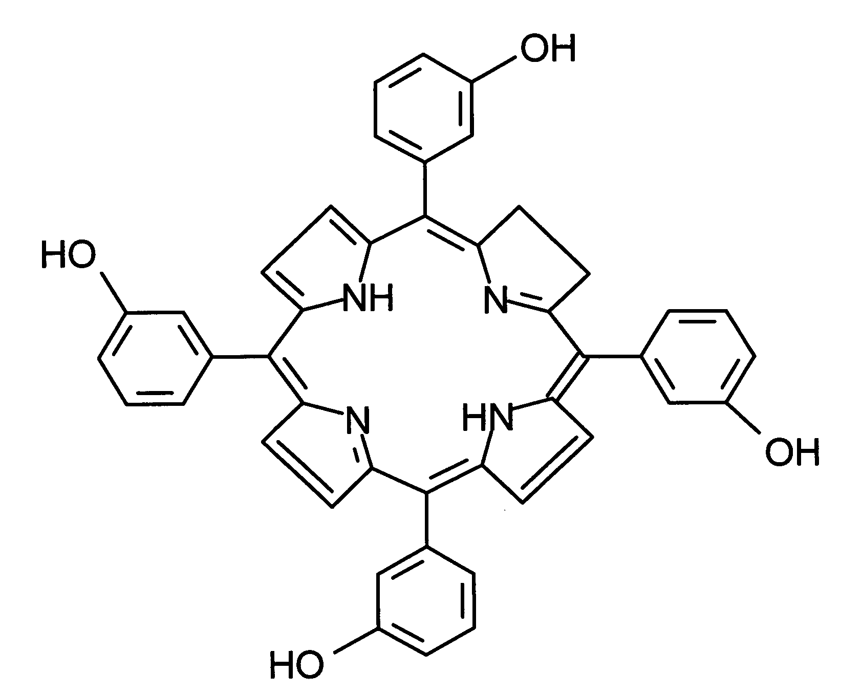
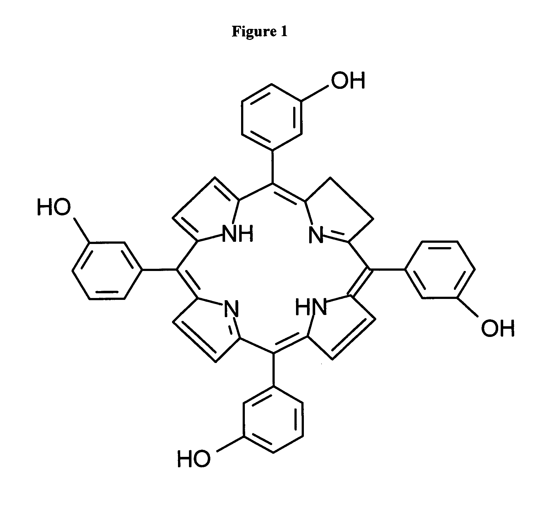
[0016]Briefly stated, in the present invention photosensitizers are used as fluorescent markers for in vivo detection of the distribution of the injected filler material during cosmetic treatments. In one preferred embodiment, liposomal formulated temoporfin is used, as the photosensitive component, in very small concentrations along with fillers for cosmetic and wound healing applications. Fillers, which can be used in the invention, include collagen, hyaluronic acids and other synthetic or natural products which are generally used in wound healing, scar reduction and other such medical applications. In another preferred embodiment the formulated photosensitizer is coupled to the filler so that tracking of the filler is possible over longer periods of time. A liposomal formulated photosensitizer is injected with the fillers into the treatment area, and is irradiated with laser light shortly after injection. The emitted fluorescence is measured by a special non-invasive device. Thereby it is possible to monitor the injection site and the distribution of the injected solution around the injection site. When irradiated with laser or other light source, the fluorescence of the photosensitizer is detected using a fluorescence detector, which permits to tracking the filler at injection site and in the injection volume.

Problems solved by technology

But unprocessed collagen is mechanically weak and vulnerable to chemical and enzymatic attacks that limits its use.

Method used


Image

  • Formulations for cosmetic and wound care treatments with photosensitizes as fluorescent markers
  • Formulations for cosmetic and wound care treatments with photosensitizes as fluorescent markers

Examples


example 1

Uses of Liposomal Temoporfin as Marker with Collagen as Filler for Wrinkle Removal

[0032]A low concentration of liposomal formulated temoporfin (3-5 μg / ml mTHPC) is injected with collagen into a treatment site. The liposome formulation of hydrophobic temoporfin is beneficial as it increases water solubility. After injection the site is illuminated with suitable excitation wavelength, which generally is different from the photosensitizer's main absorption peak, thus avoiding cytotoxic damages to cells. For temoporfin (mTHPC) its spectrum has an excitation maximum at 417 nm while the main absorption peak is at 652 nm. The photosensitizer used here serves as a visual marker indicating the success of injected collagen material.

example 2

Uses of Temoporfin as Marker to Study the Photochemical Cross-Linking of Collagen

[0033]To trace the photochemical cross-linking of collagen using photoactive compound a small amount of liposomal formulation containing a low concentration (3-15 μg / ml) of temoporfin (mTHPC) is used with collagen. In this case temoporfin is used as marker as well as photo therapeutic compound. The spectral character of temoporfin shows excitation maxima at 417 nm and an activating peak at 652 nm. Photochemical cross-linking of the collagen can be followed by monitoring the fluorescence of temoporfin excited by 417 nm.

Pepsin Promoted Collagen Gel Degradation Study Using Temoporfin Marker Material:

[0034]3 batches of collagen gels, collagen content 9.37 mg / ml Collagen I rat tail (2 test plates containing 4 samples)

TABLE 1Collagen Gel Batch FormulationIrradiation 652 nmbatchConsistencemTHPC (μg / ml)(J / cm2)1Gel0102Gel5103Gel5—

[0035]Batches 1, 2 and 3 are prepared, each containing a 15 ml solution of commerc...

example 3

Uses of Temoporfin as Marker with Hyaluronic Acid as Dermal Filler

[0038]Medical devices composed of hyaluronic acid and liposomal formulated mTHPC are used as dermal fillers. In an example, 1 ml (20 mg / ml injecting solution) of hyaluronic acid is mixed with a liposomal formulated temoporfin, wherein, temoporfin is present in a low concentration (3-10 μg / ml ) for tracking the hyaluronic acid in vivo. This formulation is water soluble and is further diluted with water to get the final injecting solution. After injecting the formulation having hyaluronic acid with a liposomal formulated temoporfin, the site is illuminated with suitable excitation wavelength which is not generally identical with main absorption (activation) peak of the temoporfin, thus avoiding cytotoxic damages to cells. The injected site may also be covered with a light blocking plaster or plastic to protect the skin area from phototoxic effect of light exposure for few days. The light was incident uniformly over the ...


PUM

PropertyMeasurementUnit
concentrationaaaaaaaaaa

Abstract

Photoactive materials, such as photosensitizers, are used as fluorescent markers for in vivo detection of the distribution of the injected filler material during cosmetic treatments. In one preferred embodiment, liposomal formulated temoporfin is used, as the photoactive component, in very small concentrations along with fillers for cosmetic and wound healing applications. Fillers, which can be used in the invention, include collagen, hyaluronic acids and other synthetic or natural products which are generally used in wound healing, scar reduction and other such medical applications. In a preferred embodiment, the formulated photosensitizer is coupled to the filler so that tracking is possible over longer periods of time A liposomal formulated photosensitizer is injected with the fillers into the treatment area, and is irradiated with laser light shortly after injection. The emitted fluorescence is measured by a special non-invasive device. Thereby it is possible to monitor the injection site and the distribution of the injected solution around the injection site. When irradiated with laser or other light source, the fluorescence of the photosensitizer is detected using a fluorescence detector, which permits tracking the filler at injection site and in the injection volume.

Description

BACKGROUND OF THE INVENTION[0001]1. Field of the Invention[0002]Present invention relates to fluorescent markers in general and more specifically for use of Photosensitizer in biological filler as fluorescent marker, to trace the success of different types of fillers used for tissue repair, and other cosmetic applications.[0003]2. Invention Disclosure Statement[0004]In cosmetic surgery and wound healing treatments natural or synthetic biological materials are used. To treat third degree burn injuries, deep cut, or even for cosmetic correction of skin imperfection biological materials called fillers are used widely. The fillers include collagen, hyaluronic acid or others such synthetic materials.[0005]The protein collagen is the main substance of connective tissue and is present in humans. In mammals collagen is the most abundant protein. Collagen gives many different organs and tissues support and elastic properties. It has been found in many different tissues and organs like bones,...

Claims


Application Information

Patent Timeline
Patent Type & Authority Applications(United States)
IPC IPC(8): A61K9/127A61K38/00A61P17/00A61K31/715
CPCA61K9/0019A61L27/50A61K31/715A61K9/06A61P17/00
Inventor NEUBERGER, WOLFGANGGRAFE, SUSANNANIFANTIEV, NIKOLAY E.
Owner CERAMOPTEC IND INC
Learn More